

6 Diagnostic Capability of Optical Coherence Tomography …
145
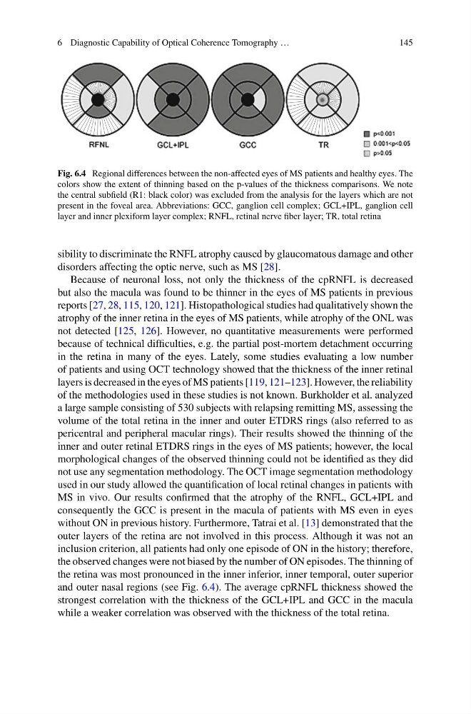
Fig. 6.4 Regional differences between the non-affected eyes of MS patients and healthy eyes. The
colors show the extent of thinning based on the p-values of the thickness comparisons. We note
the central subfield (R1: black color) was excluded from the analysis for the layers which are not
present in the foveal area. Abbreviations: GCC, ganglion cell complex; GCL+IPL, ganglion cell
layer and inner plexiform layer complex; RNFL, retinal nerve fiber layer; TR, total retina
sibility to discriminate the RNFL atrophy caused by glaucomatous damage and other
disorders affecting the optic nerve, such as MS [28].
Because of neuronal loss, not only the thickness of the cpRNFL is decreased
but also the macula was found to be thinner in the eyes of MS patients in previous
reports [27, 28, 115, 120, 121]. Histopathological studies had qualitatively shown the
atrophy of the inner retina in the eyes of MS patients, while atrophy of the ONL was
not detected [125, 126]. However, no quantitative measurements were performed
because of technical difficulties, e.g. the partial post-mortem detachment occurring
in the retina in many of the eyes. Lately, some studies evaluating a low number
of patients and using OCT technology showed that the thickness of the inner retinal
layers is decreased in the eyes of MS patients [119, 121–123]. However, the reliability
of the methodologies used in these studies is not known. Burkholder et al. analyzed
a large sample consisting of 530 subjects with relapsing remitting MS, assessing the
volume of the total retina in the inner and outer ETDRS rings (also referred to as
pericentral and peripheral macular rings). Their results showed the thinning of the
inner and outer retinal ETDRS rings in the eyes of MS patients; however, the local
morphological changes of the observed thinning could not be identified as they did
not use any segmentation methodology. The OCT image segmentation methodology
used in our study allowed the quantification of local retinal changes in patients with
MS in vivo. Our results confirmed that the atrophy of the RNFL, GCL+IPL and
consequently the GCC is present in the macula of patients with MS even in eyes
without ON in previous history. Furthermore, Tatrai et al. [13] demonstrated that the
outer layers of the retina are not involved in this process. Although it was not an
inclusion criterion, all patients had only one episode of ON in the history; therefore,
the observed changes were not biased by the number of ON episodes. The thinning of
the retina was most pronounced in the inner inferior, inner temporal, outer superior
and outer nasal regions (see Fig. 6.4). The average cpRNFL thickness showed the
strongest correlation with the thickness of the GCL+IPL and GCC in the macula
while a weaker correlation was observed with the thickness of the total retina.
145
Fig. 6.4 Regional differences between the non-affected eyes of MS patients and healthy eyes. The
colors show the extent of thinning based on the p-values of the thickness comparisons. We note
the central subfield (R1: black color) was excluded from the analysis for the layers which are not
present in the foveal area. Abbreviations: GCC, ganglion cell complex; GCL+IPL, ganglion cell
layer and inner plexiform layer complex; RNFL, retinal nerve fiber layer; TR, total retina
sibility to discriminate the RNFL atrophy caused by glaucomatous damage and other
disorders affecting the optic nerve, such as MS [28].
Because of neuronal loss, not only the thickness of the cpRNFL is decreased
but also the macula was found to be thinner in the eyes of MS patients in previous
reports [27, 28, 115, 120, 121]. Histopathological studies had qualitatively shown the
atrophy of the inner retina in the eyes of MS patients, while atrophy of the ONL was
not detected [125, 126]. However, no quantitative measurements were performed
because of technical difficulties, e.g. the partial post-mortem detachment occurring
in the retina in many of the eyes. Lately, some studies evaluating a low number
of patients and using OCT technology showed that the thickness of the inner retinal
layers is decreased in the eyes of MS patients [119, 121–123]. However, the reliability
of the methodologies used in these studies is not known. Burkholder et al. analyzed
a large sample consisting of 530 subjects with relapsing remitting MS, assessing the
volume of the total retina in the inner and outer ETDRS rings (also referred to as
pericentral and peripheral macular rings). Their results showed the thinning of the
inner and outer retinal ETDRS rings in the eyes of MS patients; however, the local
morphological changes of the observed thinning could not be identified as they did
not use any segmentation methodology. The OCT image segmentation methodology
used in our study allowed the quantification of local retinal changes in patients with
MS in vivo. Our results confirmed that the atrophy of the RNFL, GCL+IPL and
consequently the GCC is present in the macula of patients with MS even in eyes
without ON in previous history. Furthermore, Tatrai et al. [13] demonstrated that the
outer layers of the retina are not involved in this process. Although it was not an
inclusion criterion, all patients had only one episode of ON in the history; therefore,
the observed changes were not biased by the number of ON episodes. The thinning of
the retina was most pronounced in the inner inferior, inner temporal, outer superior
and outer nasal regions (see Fig. 6.4). The average cpRNFL thickness showed the
strongest correlation with the thickness of the GCL+IPL and GCC in the macula
while a weaker correlation was observed with the thickness of the total retina.