

51
(prostate cancer), A375 (malignant melanoma), HepG2 (hepatocellular cancer),
MCF-7 (breast cancer), H413 (oral carcinoma), and OKF6 (normal oral keratinocytes). The deep eutectic solvents, at different dilutions in culture medium, were
compared with their individual components using the 3-(4,5-dimethylthiazol-2-yl)2,5-diphenyltetrazolium bromide (MTT) reduction assay. The results showed appreciable toxicity of the solvents, which was generally intermediate between that of the
components, except for those based on urea, which were less toxic than the constituent chemicals. When the toxicity toward the cancer cell lines was compared with
that toward the normal keratinocytes, only a small degree of selectivity toward
malignant cells was observed.
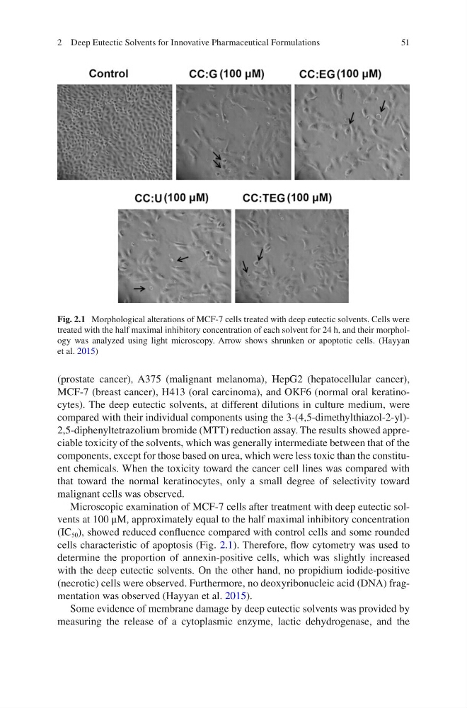
Microscopic examination of MCF-7 cells after treatment with deep eutectic solvents at 100 μM, approximately equal to the half maximal inhibitory concentration
(IC 50 ), showed reduced confluence compared with control cells and some rounded
cells characteristic of apoptosis (Fig. 2.1). Therefore, flow cytometry was used to
determine the proportion of annexin-positive cells, which was slightly increased
with the deep eutectic solvents. On the other hand, no propidium iodide-positive
(necrotic) cells were observed. Furthermore, no deoxyribonucleic acid (DNA) fragmentation was observed (Hayyan et al. 2015).
Some evidence of membrane damage by deep eutectic solvents was provided by
measuring the release of a cytoplasmic enzyme, lactic dehydrogenase, and the
Fig. 2.1 Morphological alterations of MCF-7 cells treated with deep eutectic solvents. Cells were
treated with the half maximal inhibitory concentration of each solvent for 24 h, and their morphology was analyzed using light microscopy. Arrow shows shrunken or apoptotic cells. (Hayyan
et al. 2015)
2 Deep Eutectic Solvents for Innovative Pharmaceutical Formulations
(prostate cancer), A375 (malignant melanoma), HepG2 (hepatocellular cancer),
MCF-7 (breast cancer), H413 (oral carcinoma), and OKF6 (normal oral keratinocytes). The deep eutectic solvents, at different dilutions in culture medium, were
compared with their individual components using the 3-(4,5-dimethylthiazol-2-yl)2,5-diphenyltetrazolium bromide (MTT) reduction assay. The results showed appreciable toxicity of the solvents, which was generally intermediate between that of the
components, except for those based on urea, which were less toxic than the constituent chemicals. When the toxicity toward the cancer cell lines was compared with
that toward the normal keratinocytes, only a small degree of selectivity toward
malignant cells was observed.
Microscopic examination of MCF-7 cells after treatment with deep eutectic solvents at 100 μM, approximately equal to the half maximal inhibitory concentration
(IC 50 ), showed reduced confluence compared with control cells and some rounded
cells characteristic of apoptosis (Fig. 2.1). Therefore, flow cytometry was used to
determine the proportion of annexin-positive cells, which was slightly increased
with the deep eutectic solvents. On the other hand, no propidium iodide-positive
(necrotic) cells were observed. Furthermore, no deoxyribonucleic acid (DNA) fragmentation was observed (Hayyan et al. 2015).
Some evidence of membrane damage by deep eutectic solvents was provided by
measuring the release of a cytoplasmic enzyme, lactic dehydrogenase, and the
Fig. 2.1 Morphological alterations of MCF-7 cells treated with deep eutectic solvents. Cells were
treated with the half maximal inhibitory concentration of each solvent for 24 h, and their morphology was analyzed using light microscopy. Arrow shows shrunken or apoptotic cells. (Hayyan
et al. 2015)
2 Deep Eutectic Solvents for Innovative Pharmaceutical Formulations