

limit by help of optical near-fields that are generated around single plasmonic
nanostructures and afterwards scattered to a detector [367–371]. There exists
considerable interest in combining ultrafast vibrational spectroscopy with so-called
scattering scanning near-field optical microscopy (s-SNOM) [372].
Combinations of 2D IR and s-SNOM methods have up to now not been realized.
However, the combination of metal tips and ultrafast IR spectroscopy has been
demonstrated recently as feasible and yields chemically-specific vibrational
dynamics from nanometer length scales (nanoscopy) [31, 373, 374]. The spatial
resolution of this method can be as good as 20 nm [374]. In proposed ultrafast
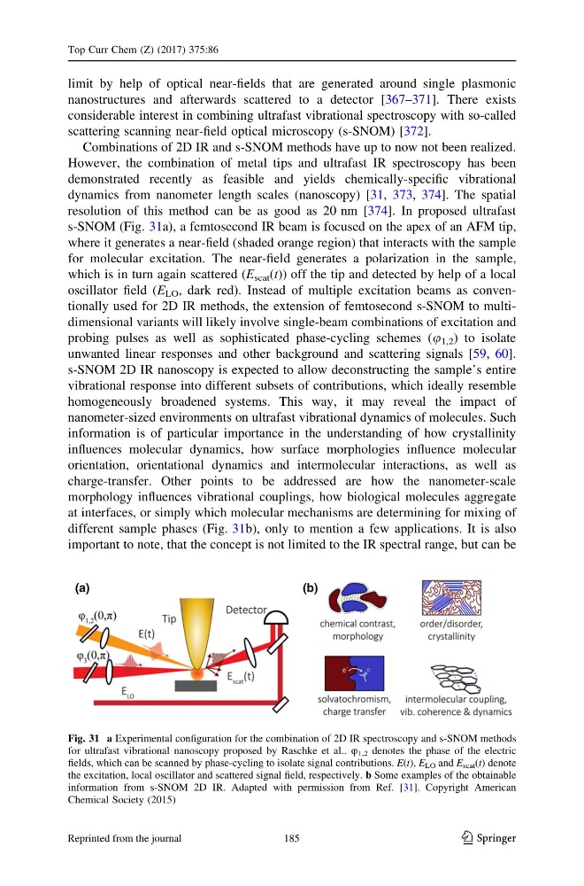
s-SNOM (Fig. 31a), a femtosecond IR beam is focused on the apex of an AFM tip,
where it generates a near-field (shaded orange region) that interacts with the sample
for molecular excitation. The near-field generates a polarization in the sample,
which is in turn again scattered (E scat (t)) off the tip and detected by help of a local
oscillator field (E LO , dark red). Instead of multiple excitation beams as conventionally used for 2D IR methods, the extension of femtosecond s-SNOM to multidimensional variants will likely involve single-beam combinations of excitation and
probing pulses as well as sophisticated phase-cycling schemes (u 1,2 ) to isolate
unwanted linear responses and other background and scattering signals [59, 60].
s-SNOM 2D IR nanoscopy is expected to allow deconstructing the sample’s entire
vibrational response into different subsets of contributions, which ideally resemble
homogeneously broadened systems. This way, it may reveal the impact of
nanometer-sized environments on ultrafast vibrational dynamics of molecules. Such
information is of particular importance in the understanding of how crystallinity
influences molecular dynamics, how surface morphologies influence molecular
orientation, orientational dynamics and intermolecular interactions, as well as
charge-transfer. Other points to be addressed are how the nanometer-scale
morphology influences vibrational couplings, how biological molecules aggregate
at interfaces, or simply which molecular mechanisms are determining for mixing of
different sample phases (Fig. 31b), only to mention a few applications. It is also
important to note, that the concept is not limited to the IR spectral range, but can be
Fig. 31 a Experimental configuration for the combination of 2D IR spectroscopy and s-SNOM methods
for ultrafast vibrational nanoscopy proposed by Raschke et al.. u 1,2 denotes the phase of the electric
fields, which can be scanned by phase-cycling to isolate signal contributions. E(t), E LO and E scat (t) denote
the excitation, local oscillator and scattered signal field, respectively. b Some examples of the obtainable
information from s-SNOM 2D IR. Adapted with permission from Ref. [31]. Copyright American
Chemical Society (2015)
Top Curr Chem (Z) (2017) 375:86
123
185
Reprinted from the journal
nanostructures and afterwards scattered to a detector [367–371]. There exists
considerable interest in combining ultrafast vibrational spectroscopy with so-called
scattering scanning near-field optical microscopy (s-SNOM) [372].
Combinations of 2D IR and s-SNOM methods have up to now not been realized.
However, the combination of metal tips and ultrafast IR spectroscopy has been
demonstrated recently as feasible and yields chemically-specific vibrational
dynamics from nanometer length scales (nanoscopy) [31, 373, 374]. The spatial
resolution of this method can be as good as 20 nm [374]. In proposed ultrafast
s-SNOM (Fig. 31a), a femtosecond IR beam is focused on the apex of an AFM tip,
where it generates a near-field (shaded orange region) that interacts with the sample
for molecular excitation. The near-field generates a polarization in the sample,
which is in turn again scattered (E scat (t)) off the tip and detected by help of a local
oscillator field (E LO , dark red). Instead of multiple excitation beams as conventionally used for 2D IR methods, the extension of femtosecond s-SNOM to multidimensional variants will likely involve single-beam combinations of excitation and
probing pulses as well as sophisticated phase-cycling schemes (u 1,2 ) to isolate
unwanted linear responses and other background and scattering signals [59, 60].
s-SNOM 2D IR nanoscopy is expected to allow deconstructing the sample’s entire
vibrational response into different subsets of contributions, which ideally resemble
homogeneously broadened systems. This way, it may reveal the impact of
nanometer-sized environments on ultrafast vibrational dynamics of molecules. Such
information is of particular importance in the understanding of how crystallinity
influences molecular dynamics, how surface morphologies influence molecular
orientation, orientational dynamics and intermolecular interactions, as well as
charge-transfer. Other points to be addressed are how the nanometer-scale
morphology influences vibrational couplings, how biological molecules aggregate
at interfaces, or simply which molecular mechanisms are determining for mixing of
different sample phases (Fig. 31b), only to mention a few applications. It is also
important to note, that the concept is not limited to the IR spectral range, but can be
Fig. 31 a Experimental configuration for the combination of 2D IR spectroscopy and s-SNOM methods
for ultrafast vibrational nanoscopy proposed by Raschke et al.. u 1,2 denotes the phase of the electric
fields, which can be scanned by phase-cycling to isolate signal contributions. E(t), E LO and E scat (t) denote
the excitation, local oscillator and scattered signal field, respectively. b Some examples of the obtainable
information from s-SNOM 2D IR. Adapted with permission from Ref. [31]. Copyright American
Chemical Society (2015)
Top Curr Chem (Z) (2017) 375:86
123
185
Reprinted from the journal