

report on coherent and incoherent energy transfer among the coupled vibrational
modes.
Experimentally, 2D VE is realized in the 2D pump-probe geometry to facilitate
phase-matching and avoid to scan separately for rephasing and non-rephasing
signals to obtain fully absorptive spectra. The signal field emission is intrinsically
phase-matched with the probe pulse and thus ‘‘self-heterodyned’’. Mechanistically,
signal contributions are different as compared to the response pathways for 2D IR
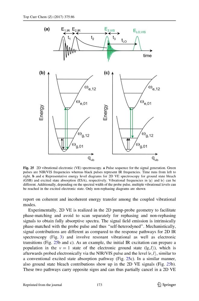
spectroscopy (Fig. 3) and involve resonant vibrational as well as electronic
transitions (Fig. 25b and c). As an example, the initial IR excitation can prepare a
population in the v = 1 state of the electronic ground state (|g,1i), which is
afterwards probed electronically via the NIR/VIS pulse and the level |e,1i, similar to
a conventional excited state absorption pathway (Fig. 25c). In a similar manner,
also ground state bleach contributions show up in the 2D VE signals (Fig. 25b).
These two pathways carry opposite signs and can thus partially cancel in a 2D VE
E 3,VIS
time
E LO,VIS
t LO
t 1
t 2
E 1,IR E 2,IR
t 3
(a)
(b)
(c)
Fig. 25 2D vibrational electronic (VE) spectroscopy. a Pulse sequence for the signal generation. Green
pulses are NIR/VIS frequencies whereas black pulses represent IR frequencies. Time runs from left to
right. b and c Representative energy level diagrams for 2D VE spectroscopy for ground state bleach
(GSB) and excited state absorption (ESA), respectively. Vibrational frequencies in |gi and |ei can be
different. Additionally, depending on the spectral width of the probe pulse, multiple vibrational levels can
be reached in the excited electronic state. Only non-rephasing diagrams are shown
Top Curr Chem (Z) (2017) 375:86
123
173
Reprinted from the journal
modes.
Experimentally, 2D VE is realized in the 2D pump-probe geometry to facilitate
phase-matching and avoid to scan separately for rephasing and non-rephasing
signals to obtain fully absorptive spectra. The signal field emission is intrinsically
phase-matched with the probe pulse and thus ‘‘self-heterodyned’’. Mechanistically,
signal contributions are different as compared to the response pathways for 2D IR
spectroscopy (Fig. 3) and involve resonant vibrational as well as electronic
transitions (Fig. 25b and c). As an example, the initial IR excitation can prepare a
population in the v = 1 state of the electronic ground state (|g,1i), which is
afterwards probed electronically via the NIR/VIS pulse and the level |e,1i, similar to
a conventional excited state absorption pathway (Fig. 25c). In a similar manner,
also ground state bleach contributions show up in the 2D VE signals (Fig. 25b).
These two pathways carry opposite signs and can thus partially cancel in a 2D VE
E 3,VIS
time
E LO,VIS
t LO
t 1
t 2
E 1,IR E 2,IR
t 3
(a)
(b)
(c)
Fig. 25 2D vibrational electronic (VE) spectroscopy. a Pulse sequence for the signal generation. Green
pulses are NIR/VIS frequencies whereas black pulses represent IR frequencies. Time runs from left to
right. b and c Representative energy level diagrams for 2D VE spectroscopy for ground state bleach
(GSB) and excited state absorption (ESA), respectively. Vibrational frequencies in |gi and |ei can be
different. Additionally, depending on the spectral width of the probe pulse, multiple vibrational levels can
be reached in the excited electronic state. Only non-rephasing diagrams are shown
Top Curr Chem (Z) (2017) 375:86
123
173
Reprinted from the journal