

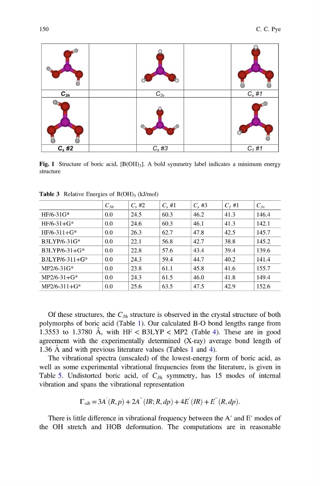
Of these structures, the C 3h structure is observed in the crystal structure of both
polymorphs of boric acid (Table 1). Our calculated B-O bond lengths range from
1.3553 to 1.3780 Å, with HF < B3LYP < MP2 (Table 4). These are in good
agreement with the experimentally determined (X-ray) average bond length of
1.36 Å and with previous literature values (Tables 1 and 4).
The vibrational spectra (unscaled) of the lowest-energy form of boric acid, as
well as some experimental vibrational frequencies from the literature, is given in
Table 5. Undistorted boric acid, of C 3h symmetry, has 15 modes of internal
vibration and spans the vibrational representation
Γ vib = 3A
′ R, p
ð
Þ+ 2A
′′ IR; R, dp
ð
Þ+ 4E
′ IR
ð Þ+ E
′′ R, dp
ð
Þ.
There is little difference in vibrational frequency between the A′ and E′ modes of
the OH stretch and HOB deformation. The computations are in reasonable
C 3h
C 3v
C s #1
C s #2
C s
C
3
#
1 #1
Fig. 1 Structure of boric acid, [B(OH) 3 ]. A bold symmetry label indicates a minimum energy
structure
Table 3 Relative Energies of B(OH) 3 (kJ/mol)
C 3h
C s #2
C s #1
C s #3
C 1 #1
C 3v
HF/6-31G*
0.0
24.5
60.3
46.2
41.3
146.4
HF/6-31+G*
0.0
24.6
60.3
46.1
41.3
142.1
HF/6-311+G*
0.0
26.3
62.7
47.8
42.5
145.7
B3LYP/6-31G*
0.0
22.1
56.8
42.7
38.8
145.2
B3LYP/6-31+G*
0.0
22.8
57.6
43.4
39.4
139.6
B3LYP/6-311+G*
0.0
24.3
59.4
44.7
40.2
141.4
MP2/6-31G*
0.0
23.8
61.1
45.8
41.6
155.7
MP2/6-31+G*
0.0
24.3
61.5
46.0
41.8
149.4
MP2/6-311+G*
0.0
25.6
63.5
47.5
42.9
152.6
150
C. C. Pye
polymorphs of boric acid (Table 1). Our calculated B-O bond lengths range from
1.3553 to 1.3780 Å, with HF < B3LYP < MP2 (Table 4). These are in good
agreement with the experimentally determined (X-ray) average bond length of
1.36 Å and with previous literature values (Tables 1 and 4).
The vibrational spectra (unscaled) of the lowest-energy form of boric acid, as
well as some experimental vibrational frequencies from the literature, is given in
Table 5. Undistorted boric acid, of C 3h symmetry, has 15 modes of internal
vibration and spans the vibrational representation
Γ vib = 3A
′ R, p
ð
Þ+ 2A
′′ IR; R, dp
ð
Þ+ 4E
′ IR
ð Þ+ E
′′ R, dp
ð
Þ.
There is little difference in vibrational frequency between the A′ and E′ modes of
the OH stretch and HOB deformation. The computations are in reasonable
C 3h
C 3v
C s #1
C s #2
C s
C
3
#
1 #1
Fig. 1 Structure of boric acid, [B(OH) 3 ]. A bold symmetry label indicates a minimum energy
structure
Table 3 Relative Energies of B(OH) 3 (kJ/mol)
C 3h
C s #2
C s #1
C s #3
C 1 #1
C 3v
HF/6-31G*
0.0
24.5
60.3
46.2
41.3
146.4
HF/6-31+G*
0.0
24.6
60.3
46.1
41.3
142.1
HF/6-311+G*
0.0
26.3
62.7
47.8
42.5
145.7
B3LYP/6-31G*
0.0
22.1
56.8
42.7
38.8
145.2
B3LYP/6-31+G*
0.0
22.8
57.6
43.4
39.4
139.6
B3LYP/6-311+G*
0.0
24.3
59.4
44.7
40.2
141.4
MP2/6-31G*
0.0
23.8
61.1
45.8
41.6
155.7
MP2/6-31+G*
0.0
24.3
61.5
46.0
41.8
149.4
MP2/6-311+G*
0.0
25.6
63.5
47.5
42.9
152.6
150
C. C. Pye