

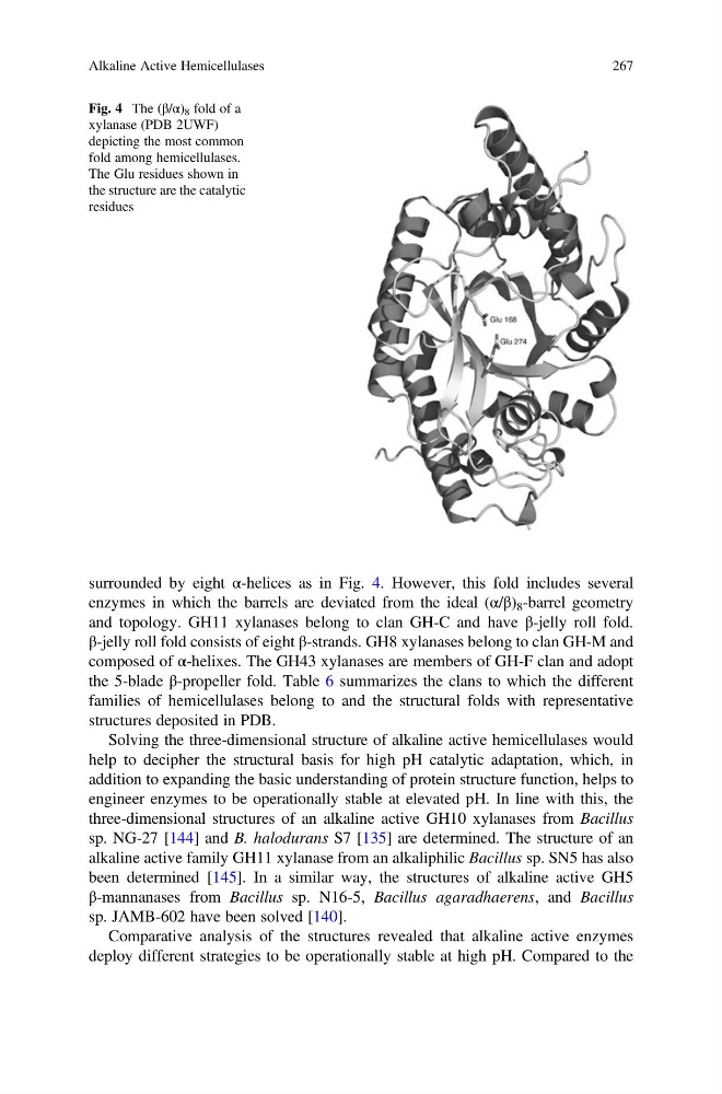
surrounded by eight α-helices as in Fig. 4. However, this fold includes several
enzymes in which the barrels are deviated from the ideal (α/β) 8 -barrel geometry
and topology. GH11 xylanases belong to clan GH-C and have β-jelly roll fold.
β-jelly roll fold consists of eight β-strands. GH8 xylanases belong to clan GH-M and
composed of α-helixes. The GH43 xylanases are members of GH-F clan and adopt
the 5-blade β-propeller fold. Table 6 summarizes the clans to which the different
families of hemicellulases belong to and the structural folds with representative
structures deposited in PDB.
Solving the three-dimensional structure of alkaline active hemicellulases would
help to decipher the structural basis for high pH catalytic adaptation, which, in
addition to expanding the basic understanding of protein structure function, helps to
engineer enzymes to be operationally stable at elevated pH. In line with this, the
three-dimensional structures of an alkaline active GH10 xylanases from Bacillus
sp. NG-27 [144] and B. halodurans S7 [135] are determined. The structure of an
alkaline active family GH11 xylanase from an alkaliphilic Bacillus sp. SN5 has also
been determined [145]. In a similar way, the structures of alkaline active GH5
β-mannanases from Bacillus sp. N16-5, Bacillus agaradhaerens, and Bacillus
sp. JAMB-602 have been solved [140].
Comparative analysis of the structures revealed that alkaline active enzymes
deploy different strategies to be operationally stable at high pH. Compared to the
Fig. 4 The (β/α) 8 fold of a
xylanase (PDB 2UWF)
depicting the most common
fold among hemicellulases.
The Glu residues shown in
the structure are the catalytic
residues
Alkaline Active Hemicellulases
267
enzymes in which the barrels are deviated from the ideal (α/β) 8 -barrel geometry
and topology. GH11 xylanases belong to clan GH-C and have β-jelly roll fold.
β-jelly roll fold consists of eight β-strands. GH8 xylanases belong to clan GH-M and
composed of α-helixes. The GH43 xylanases are members of GH-F clan and adopt
the 5-blade β-propeller fold. Table 6 summarizes the clans to which the different
families of hemicellulases belong to and the structural folds with representative
structures deposited in PDB.
Solving the three-dimensional structure of alkaline active hemicellulases would
help to decipher the structural basis for high pH catalytic adaptation, which, in
addition to expanding the basic understanding of protein structure function, helps to
engineer enzymes to be operationally stable at elevated pH. In line with this, the
three-dimensional structures of an alkaline active GH10 xylanases from Bacillus
sp. NG-27 [144] and B. halodurans S7 [135] are determined. The structure of an
alkaline active family GH11 xylanase from an alkaliphilic Bacillus sp. SN5 has also
been determined [145]. In a similar way, the structures of alkaline active GH5
β-mannanases from Bacillus sp. N16-5, Bacillus agaradhaerens, and Bacillus
sp. JAMB-602 have been solved [140].
Comparative analysis of the structures revealed that alkaline active enzymes
deploy different strategies to be operationally stable at high pH. Compared to the
Fig. 4 The (β/α) 8 fold of a
xylanase (PDB 2UWF)
depicting the most common
fold among hemicellulases.
The Glu residues shown in
the structure are the catalytic
residues
Alkaline Active Hemicellulases
267