

substitution of Lys by Arg may allow formation of hydrogen bonds at extremely
high pH. His and Gln are largely neutral at alkaline condition, and this may be
important to maintain the protein solubility in the alkaline condition. Asn is one of
the most alkali susceptible residues [210, 211], and hence, its occurrence in alkalineadapted proteins, especially on exposed surfaces, is relatively low [206]. This agrees
to previous studies that involved mutational substitution of Asn with less susceptible
amino acids and resulted in a better stability at high pH [212, 213].
Charged residues are known to play important roles in structural adaptation of
biomolecules. Such residues are vital in high pH adaptations. Extracellular products
of alkaliphiles tend to have more acidic residues on their surfaces than their
non-alkaliphilic counterparts. For instance, deduced amino acid sequence analysis
has shown that the externally exposed alkaliphile membrane protein loops have
acidic residues, while the non-alkaliphile homologue loops have neutral/basic residues [214]. Similarly, as described above in Sect. 3.2, the surface exposed proteins
and polysaccharides of alkaliphiles such as SplA, liposaccharides, and SCWPs are
rich in negatively charged residues. Structural analysis of extracellular enzymes also
reveals that their surface is more acidic than that of non-alkaline active counterparts.
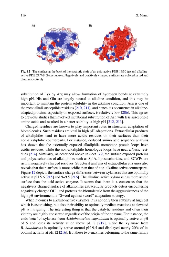
Figure 12 depicts the surface charge difference between xylanases that are optimally
active at pH 5.6 [215] and 9–9.5 [216]. The alkaline active xylanase has more acidic
surface than the acid-active enzyme. It seems that there is a consensus that the
negatively charged surface of alkaliphiles extracellular products deters encountering
negatively charged OH
À and protects the biomolecule from the aggressiveness of the
high pH environment, a “Sword against sword” adaptation strategy.
When it comes to alkaline-active enzymes, it is not only their stability at high pH
which is astonishing, but also their ability to optimally mediate reactions at elevated
pH is intriguing. The interesting thing is that the catalytic residues and often their
vicinity are highly conserved regardless of the origin of the enzyme. For instance, the
endo-beta-1,4-xylanase from Acidobacterium capsulatum is optimally active at pH
of 5 and loses its activity at or above pH 8 [217], while the xylanase from
B. halodurans is optimally active around pH 9.5 and displayed nearly 20% of its
optimal activity at pH 12 [216]. But these two enzymes belonging to the same family
Fig. 12 The surface at the back of the catalytic cleft of an acid-active PDB 1B30 (a) and alkalineactive PDB 2UWF (b) xylanases. Negatively and positively charged surfaces are colored in red and
blue, respectively
116
G. Mamo
high pH. His and Gln are largely neutral at alkaline condition, and this may be
important to maintain the protein solubility in the alkaline condition. Asn is one of
the most alkali susceptible residues [210, 211], and hence, its occurrence in alkalineadapted proteins, especially on exposed surfaces, is relatively low [206]. This agrees
to previous studies that involved mutational substitution of Asn with less susceptible
amino acids and resulted in a better stability at high pH [212, 213].
Charged residues are known to play important roles in structural adaptation of
biomolecules. Such residues are vital in high pH adaptations. Extracellular products
of alkaliphiles tend to have more acidic residues on their surfaces than their
non-alkaliphilic counterparts. For instance, deduced amino acid sequence analysis
has shown that the externally exposed alkaliphile membrane protein loops have
acidic residues, while the non-alkaliphile homologue loops have neutral/basic residues [214]. Similarly, as described above in Sect. 3.2, the surface exposed proteins
and polysaccharides of alkaliphiles such as SplA, liposaccharides, and SCWPs are
rich in negatively charged residues. Structural analysis of extracellular enzymes also
reveals that their surface is more acidic than that of non-alkaline active counterparts.
Figure 12 depicts the surface charge difference between xylanases that are optimally
active at pH 5.6 [215] and 9–9.5 [216]. The alkaline active xylanase has more acidic
surface than the acid-active enzyme. It seems that there is a consensus that the
negatively charged surface of alkaliphiles extracellular products deters encountering
negatively charged OH
À and protects the biomolecule from the aggressiveness of the
high pH environment, a “Sword against sword” adaptation strategy.
When it comes to alkaline-active enzymes, it is not only their stability at high pH
which is astonishing, but also their ability to optimally mediate reactions at elevated
pH is intriguing. The interesting thing is that the catalytic residues and often their
vicinity are highly conserved regardless of the origin of the enzyme. For instance, the
endo-beta-1,4-xylanase from Acidobacterium capsulatum is optimally active at pH
of 5 and loses its activity at or above pH 8 [217], while the xylanase from
B. halodurans is optimally active around pH 9.5 and displayed nearly 20% of its
optimal activity at pH 12 [216]. But these two enzymes belonging to the same family
Fig. 12 The surface at the back of the catalytic cleft of an acid-active PDB 1B30 (a) and alkalineactive PDB 2UWF (b) xylanases. Negatively and positively charged surfaces are colored in red and
blue, respectively
116
G. Mamo