

straightforward encapsulation and size control due to much simpler processing
[91]. Another feature of these charged polypeptide vesicles is the potential for
facile functionalization of the hydrophilic polypeptide chains at the vesicle surface
either through chemical conjugation to amine or carboxylate residues [93] or
through careful choice of charged residues.
Addressing this point, Deming’s laboratory reported the preparation of
arginine–leucine (i.e., R 60 L 20 ) vesicles that are able to readily enter cells due to
the many guanidinium groups of the arginine segments [94]. In this case,
the arginine residues play a dual role, being both structure-directing in vesicle
formation as well as functional for cell binding and entry. Studies on endocytotsis
and intracellular trafficking of these vesicles revealed that they enter HeLa cells
primarily via macropinocytosis [95]. They were found to primarily reside in early
endosomes, but not in lysosomes, and although some manage to escape into
cytoplasm many are trapped within these compartments. Regardless, another
study showed that R 60 L 20 vesicles were effective at condensing plasmid DNA
and transfecting it into a variety of cell lines, showing the vesicles do have potential
for intracellular delivery [96]. These DNA carriers are advantageous over many
other transfection agents due to their low cytotoxicity.
From the pioneering studies on block copolypeptide vesicles described above,
design criteria were established for successful vesicle formation, namely an
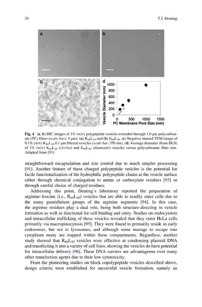
Fig. 4 (a, b) DIC images of 1% (w/v) polypeptide vesicles extruded through 1.0 μm polycarbonate (PC) filters (scale bars: 5 μm): (a) K 60 L 20 and (b) E 60 L 20 . (c) Negative stained TEM image of
0.1% (w/v) K 60 L 20 0.1 μm filtered vesicles (scale bar: 350 nm). (d) Average diameter (from DLS)
of 1% (w/v) K 60 L 20 (circles) and E 60 L 20 (diamonds) vesicles versus polycarbonate filter size.
Adapted from [91]
24
T.J. Deming
[91]. Another feature of these charged polypeptide vesicles is the potential for
facile functionalization of the hydrophilic polypeptide chains at the vesicle surface
either through chemical conjugation to amine or carboxylate residues [93] or
through careful choice of charged residues.
Addressing this point, Deming’s laboratory reported the preparation of
arginine–leucine (i.e., R 60 L 20 ) vesicles that are able to readily enter cells due to
the many guanidinium groups of the arginine segments [94]. In this case,
the arginine residues play a dual role, being both structure-directing in vesicle
formation as well as functional for cell binding and entry. Studies on endocytotsis
and intracellular trafficking of these vesicles revealed that they enter HeLa cells
primarily via macropinocytosis [95]. They were found to primarily reside in early
endosomes, but not in lysosomes, and although some manage to escape into
cytoplasm many are trapped within these compartments. Regardless, another
study showed that R 60 L 20 vesicles were effective at condensing plasmid DNA
and transfecting it into a variety of cell lines, showing the vesicles do have potential
for intracellular delivery [96]. These DNA carriers are advantageous over many
other transfection agents due to their low cytotoxicity.
From the pioneering studies on block copolypeptide vesicles described above,
design criteria were established for successful vesicle formation, namely an
Fig. 4 (a, b) DIC images of 1% (w/v) polypeptide vesicles extruded through 1.0 μm polycarbonate (PC) filters (scale bars: 5 μm): (a) K 60 L 20 and (b) E 60 L 20 . (c) Negative stained TEM image of
0.1% (w/v) K 60 L 20 0.1 μm filtered vesicles (scale bar: 350 nm). (d) Average diameter (from DLS)
of 1% (w/v) K 60 L 20 (circles) and E 60 L 20 (diamonds) vesicles versus polycarbonate filter size.
Adapted from [91]
24
T.J. Deming