

50
4
4.3.3 Voltage-Gated Ca 2+ -Channels
Voltage-gated Ca 2+ -channels include the CaV1-L-type channels that provide long- lasting
Ca 2+ - currents in the skeletal muscle, heart, neurons and endocrine cells. CaV2-N- type,
P/Q-type and R-type channels are involved in synaptic transmission in the nervous system. CaV3 T-type channels have a transient kinetics and include pacemaker channels in
sinoatrial nodes as well as channels involved in stimulus-secretion coupling in gland cells,
neuronal rhythmicity and the sperm acrosome reaction (Berridge 2012). VOC channels
open upon membrane depolarization, resulting in a cytosolic Ca 2+ -spike. Immediately
after Ca 2+ -entry, the channel is inactivated, ensuring that no more Ca 2+ can get in and the
spike diffuses. After recovery from inactivation, the channel is ready for another opening.
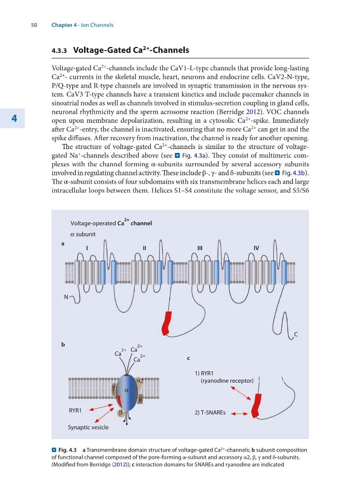
The structure of voltage-gated Ca 2+ -channels is similar to the structure of voltagegated Na + -channels described above (see . Fig. 4.3a). They consist of multimeric complexes with the channel forming α-subunits surrounded by several accessory subunits
involved in regulating channel activity. These include β-, γ- and δ-subunits (see . Fig. 4.3b).
The α-subunit consists of four subdomains with six transmembrane helices each and large
intracellular loops between them. Helices S1–S4 constitute the voltage sensor, and S5/S6
N
I
I I
III
IV
...
...
c
1) RYR1
(ryanodine receptor)
2) T-SNAREs
α subunit
RYR1
Synaptic vesicle
δ
α2
β
γ
Ca
2+ Ca
2+
Ca
2+
Voltage-operated Ca
2+ channel
C
...
...
α
a
b
. Fig. 4.3 a Transmembrane domain structure of voltage-gated Ca 2+ -channels; b subunit composition
of functional channel composed of the pore-forming α-subunit and accessory α2, β, γ and δ-subunits.
(Modified from Berridge (2012)); c interaction domains for SNAREs and ryanodine are indicated
Chapter 4 · Ion Channels
4
4.3.3 Voltage-Gated Ca 2+ -Channels
Voltage-gated Ca 2+ -channels include the CaV1-L-type channels that provide long- lasting
Ca 2+ - currents in the skeletal muscle, heart, neurons and endocrine cells. CaV2-N- type,
P/Q-type and R-type channels are involved in synaptic transmission in the nervous system. CaV3 T-type channels have a transient kinetics and include pacemaker channels in
sinoatrial nodes as well as channels involved in stimulus-secretion coupling in gland cells,
neuronal rhythmicity and the sperm acrosome reaction (Berridge 2012). VOC channels
open upon membrane depolarization, resulting in a cytosolic Ca 2+ -spike. Immediately
after Ca 2+ -entry, the channel is inactivated, ensuring that no more Ca 2+ can get in and the
spike diffuses. After recovery from inactivation, the channel is ready for another opening.
The structure of voltage-gated Ca 2+ -channels is similar to the structure of voltagegated Na + -channels described above (see . Fig. 4.3a). They consist of multimeric complexes with the channel forming α-subunits surrounded by several accessory subunits
involved in regulating channel activity. These include β-, γ- and δ-subunits (see . Fig. 4.3b).
The α-subunit consists of four subdomains with six transmembrane helices each and large
intracellular loops between them. Helices S1–S4 constitute the voltage sensor, and S5/S6
N
I
I I
III
IV
...
...
c
1) RYR1
(ryanodine receptor)
2) T-SNAREs
α subunit
RYR1
Synaptic vesicle
δ
α2
β
γ
Ca
2+ Ca
2+
Ca
2+
Voltage-operated Ca
2+ channel
C
...
...
α
a
b
. Fig. 4.3 a Transmembrane domain structure of voltage-gated Ca 2+ -channels; b subunit composition
of functional channel composed of the pore-forming α-subunit and accessory α2, β, γ and δ-subunits.
(Modified from Berridge (2012)); c interaction domains for SNAREs and ryanodine are indicated
Chapter 4 · Ion Channels