

48
4
(K v 5, 6, 8 and 9 family members) act as silencers when forming hetero-oligomers with
ion-conducting channel forming subunits (Gutman et al. 2005). For each α-subunit, the
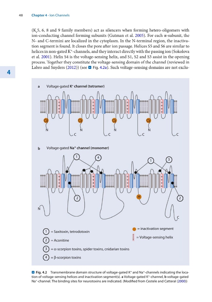
N- and C-termini are localized in the cytoplasm. In the N-terminal region, the inactivation segment is found. It closes the pore after ion passage. Helices S5 and S6 are similar to
helices in non-gated K + -channels, and they interact directly with the passing ion (Sokolova
et al. 2001). Helix S4 is the voltage-sensing helix, and S1, S2 and S3 assist in the opening
process. Together they constitute the voltage-sensing domain of the channel (reviewed in
Labro and Snyders (2012)) (see . Fig. 4.2a). Such voltage-sensing domains are not excluVoltage-gated K
+ channel (tetramer)
N
C
N
C
N
C
N
C
= inactivation segment
Voltage-gated Na + channel (monomer)
a
b
N
C
H
+
+
+
+
+
+
+
+
+
+
+
+
+
+
+
+
+
+
+
+
+
+
+
+
+
+
+
+
+
+
+
+
= Voltage-sensing helix
+
+
+
+
2
2
1 = Saxitoxin, tetrodotoxin
1
1
2 = Aconitine
3 = α-scorpion toxins, spider toxins, cnidarian toxins
4 = β-scorpion toxins
3
4
+
+
+
+
. Fig. 4.2 Transmembrane domain structure of voltage-gated K + and Na + -channels indicating the location of voltage-sensing helices and inactivation segment(s). a Voltage-gated K + -channel, b voltage-gated
Na + -channel. The binding sites for neurotoxins are indicated. (Modified from Cestele and Catteral (2000))
Chapter 4 · Ion Channels
4
(K v 5, 6, 8 and 9 family members) act as silencers when forming hetero-oligomers with
ion-conducting channel forming subunits (Gutman et al. 2005). For each α-subunit, the
N- and C-termini are localized in the cytoplasm. In the N-terminal region, the inactivation segment is found. It closes the pore after ion passage. Helices S5 and S6 are similar to
helices in non-gated K + -channels, and they interact directly with the passing ion (Sokolova
et al. 2001). Helix S4 is the voltage-sensing helix, and S1, S2 and S3 assist in the opening
process. Together they constitute the voltage-sensing domain of the channel (reviewed in
Labro and Snyders (2012)) (see . Fig. 4.2a). Such voltage-sensing domains are not excluVoltage-gated K
+ channel (tetramer)
N
C
N
C
N
C
N
C
= inactivation segment
Voltage-gated Na + channel (monomer)
a
b
N
C
H
+
+
+
+
+
+
+
+
+
+
+
+
+
+
+
+
+
+
+
+
+
+
+
+
+
+
+
+
+
+
+
+
= Voltage-sensing helix
+
+
+
+
2
2
1 = Saxitoxin, tetrodotoxin
1
1
2 = Aconitine
3 = α-scorpion toxins, spider toxins, cnidarian toxins
4 = β-scorpion toxins
3
4
+
+
+
+
. Fig. 4.2 Transmembrane domain structure of voltage-gated K + and Na + -channels indicating the location of voltage-sensing helices and inactivation segment(s). a Voltage-gated K + -channel, b voltage-gated
Na + -channel. The binding sites for neurotoxins are indicated. (Modified from Cestele and Catteral (2000))
Chapter 4 · Ion Channels