

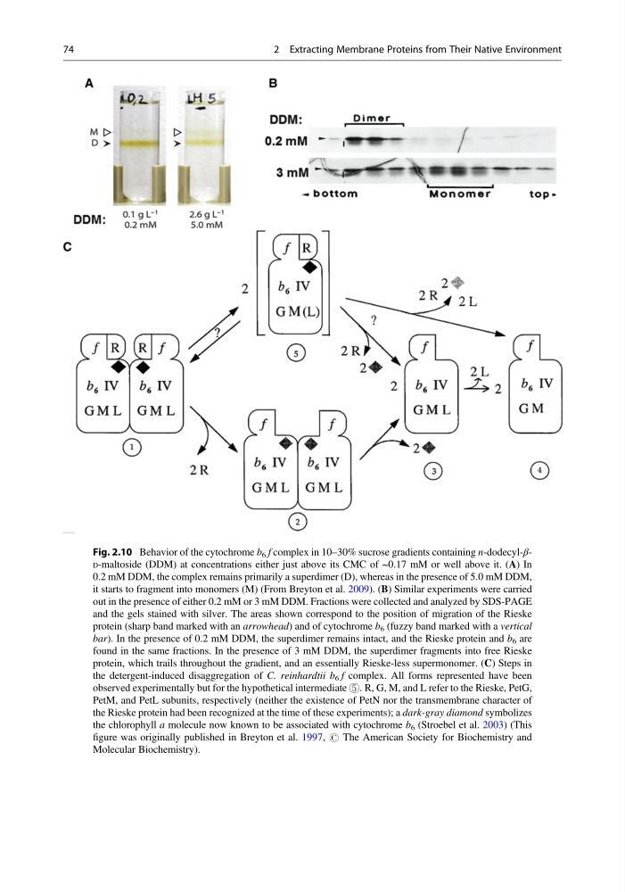
Fig. 2.10 Behavior of the cytochrome b 6 f complex in 10–30% sucrose gradients containing n-dodecyl-βD-maltoside (DDM) at concentrations either just above its CMC of ~0.17 mM or well above it. (A) In
0.2 mM DDM, the complex remains primarily a superdimer (D), whereas in the presence of 5.0 mM DDM,
it starts to fragment into monomers (M) (From Breyton et al. 2009). (B) Similar experiments were carried
out in the presence of either 0.2 mM or 3 mM DDM. Fractions were collected and analyzed by SDS-PAGE
and the gels stained with silver. The areas shown correspond to the position of migration of the Rieske
protein (sharp band marked with an arrowhead) and of cytochrome b 6 (fuzzy band marked with a vertical
bar). In the presence of 0.2 mM DDM, the superdimer remains intact, and the Rieske protein and b 6 are
found in the same fractions. In the presence of 3 mM DDM, the superdimer fragments into free Rieske
protein, which trails throughout the gradient, and an essentially Rieske-less supermonomer. (C) Steps in
the detergent-induced disaggregation of C. reinhardtii b 6 f complex. All forms represented have been
observed experimentally but for the hypothetical intermediate ⑤. R, G, M, and L refer to the Rieske, PetG,
PetM, and PetL subunits, respectively (neither the existence of PetN nor the transmembrane character of
the Rieske protein had been recognized at the time of these experiments); a dark-gray diamond symbolizes
the chlorophyll a molecule now known to be associated with cytochrome b 6 (Stroebel et al. 2003) (This
figure was originally published in Breyton et al. 1997, # The American Society for Biochemistry and
Molecular Biochemistry).
74
2 Extracting Membrane Proteins from Their Native Environment
0.2 mM DDM, the complex remains primarily a superdimer (D), whereas in the presence of 5.0 mM DDM,
it starts to fragment into monomers (M) (From Breyton et al. 2009). (B) Similar experiments were carried
out in the presence of either 0.2 mM or 3 mM DDM. Fractions were collected and analyzed by SDS-PAGE
and the gels stained with silver. The areas shown correspond to the position of migration of the Rieske
protein (sharp band marked with an arrowhead) and of cytochrome b 6 (fuzzy band marked with a vertical
bar). In the presence of 0.2 mM DDM, the superdimer remains intact, and the Rieske protein and b 6 are
found in the same fractions. In the presence of 3 mM DDM, the superdimer fragments into free Rieske
protein, which trails throughout the gradient, and an essentially Rieske-less supermonomer. (C) Steps in
the detergent-induced disaggregation of C. reinhardtii b 6 f complex. All forms represented have been
observed experimentally but for the hypothetical intermediate ⑤. R, G, M, and L refer to the Rieske, PetG,
PetM, and PetL subunits, respectively (neither the existence of PetN nor the transmembrane character of
the Rieske protein had been recognized at the time of these experiments); a dark-gray diamond symbolizes
the chlorophyll a molecule now known to be associated with cytochrome b 6 (Stroebel et al. 2003) (This
figure was originally published in Breyton et al. 1997, # The American Society for Biochemistry and
Molecular Biochemistry).
74
2 Extracting Membrane Proteins from Their Native Environment