

12.3.3 Cryo-EM Studies of MP/APol Complexes (2011–Present)
A decisive impetus was given to the use of APols for cryo-EM studies of MPs and MP complexes by
Study 12.5, carried out by the laboratory of Werner Kühlbrandt, in which supercomplex B from the
inner membrane of mitochondria was solubilized using digitonin, transferred to A8-35, purified by
sucrose gradient centrifugation, and imaged after freezing in vitreous ice (Althoff 2011; Althoff et al.
2011). Supercomplex B, the “respirasome,” comprises one copy of Complex I (NADH:ubiquinone
oxidoreductase), a superdimer of Complex III (cytochrome bc 1 ), and one copy of Complex IV
(cytochrome c oxidase), for a total mass of ~1.7 MDa. Complex I transfers electrons from NADH to
ubiquinone, Complex III from ubiquinol to cytochrome c, and Complex IV from cytochrome c to
dioxygen, the whole process generating water and a proton gradient, which the F 1 F O ATP synthase
uses for synthesizing ATP (for a review, see Kühlbrandt 2015).
Mitochondria were isolated from bovine heart tissue and supercomplexes solubilized with
digitonin. However, digitonin-solubilized supercomplexes did not partition well into the holes of
holey carbon films. Following solubilization, digitonin was therefore replaced by A8-35 using γcyclodextrin, and the various APol-trapped supercomplexes separated from one another and from free
APol by sucrose gradient ultracentrifugation in the absence of surfactant. The gradient purification
worked better than in digitonin, due to larger differences in apparent molecular mass between the
supercomplexes, which resulted in improved separation. Fractions containing APol-trapped
supercomplex B were identified on the basis of their migration in Blue-Native gels and enzymatic
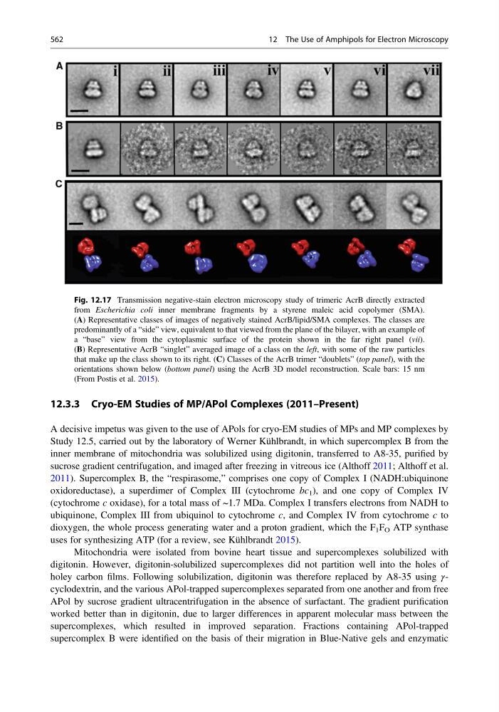
Fig. 12.17 Transmission negative-stain electron microscopy study of trimeric AcrB directly extracted
from Escherichia coli inner membrane fragments by a styrene maleic acid copolymer (SMA).
(A) Representative classes of images of negatively stained AcrB/lipid/SMA complexes. The classes are
predominantly of a “side” view, equivalent to that viewed from the plane of the bilayer, with an example of
a “base” view from the cytoplasmic surface of the protein shown in the far right panel (vii).
(B) Representative AcrB “singlet” averaged image of a class on the left, with some of the raw particles
that make up the class shown to its right. (C) Classes of the AcrB trimer “doublets” (top panel), with the
orientations shown below (bottom panel) using the AcrB 3D model reconstruction. Scale bars: 15 nm
(From Postis et al. 2015).
562
12 The Use of Amphipols for Electron Microscopy
A decisive impetus was given to the use of APols for cryo-EM studies of MPs and MP complexes by
Study 12.5, carried out by the laboratory of Werner Kühlbrandt, in which supercomplex B from the
inner membrane of mitochondria was solubilized using digitonin, transferred to A8-35, purified by
sucrose gradient centrifugation, and imaged after freezing in vitreous ice (Althoff 2011; Althoff et al.
2011). Supercomplex B, the “respirasome,” comprises one copy of Complex I (NADH:ubiquinone
oxidoreductase), a superdimer of Complex III (cytochrome bc 1 ), and one copy of Complex IV
(cytochrome c oxidase), for a total mass of ~1.7 MDa. Complex I transfers electrons from NADH to
ubiquinone, Complex III from ubiquinol to cytochrome c, and Complex IV from cytochrome c to
dioxygen, the whole process generating water and a proton gradient, which the F 1 F O ATP synthase
uses for synthesizing ATP (for a review, see Kühlbrandt 2015).
Mitochondria were isolated from bovine heart tissue and supercomplexes solubilized with
digitonin. However, digitonin-solubilized supercomplexes did not partition well into the holes of
holey carbon films. Following solubilization, digitonin was therefore replaced by A8-35 using γcyclodextrin, and the various APol-trapped supercomplexes separated from one another and from free
APol by sucrose gradient ultracentrifugation in the absence of surfactant. The gradient purification
worked better than in digitonin, due to larger differences in apparent molecular mass between the
supercomplexes, which resulted in improved separation. Fractions containing APol-trapped
supercomplex B were identified on the basis of their migration in Blue-Native gels and enzymatic
Fig. 12.17 Transmission negative-stain electron microscopy study of trimeric AcrB directly extracted
from Escherichia coli inner membrane fragments by a styrene maleic acid copolymer (SMA).
(A) Representative classes of images of negatively stained AcrB/lipid/SMA complexes. The classes are
predominantly of a “side” view, equivalent to that viewed from the plane of the bilayer, with an example of
a “base” view from the cytoplasmic surface of the protein shown in the far right panel (vii).
(B) Representative AcrB “singlet” averaged image of a class on the left, with some of the raw particles
that make up the class shown to its right. (C) Classes of the AcrB trimer “doublets” (top panel), with the
orientations shown below (bottom panel) using the AcrB 3D model reconstruction. Scale bars: 15 nm
(From Postis et al. 2015).
562
12 The Use of Amphipols for Electron Microscopy