

12.3.1 The Early Years: 1998–2008
As recalled in the introduction, the first EM study of MP/APol complexes (Study 12.1 in Table 12.1)
was a STEM study, aimed at establishing whether trapping with APols respected the dispersity of the
protein in detergent solution. The test MP, cytochrome b 6 f, was prepared in solutions of the detergent
Hecameg under two forms: an intact superdimer, comprising bound lipids, and a delipidated, lighter
form, which is a super-monomer that has lost some of the subunits present in the dimeric form (Breyton
et al. 1997). The two forms were transferred to APols A8-35 or A8-75 by mixing, dilution under the
CMC of Hecameg, and ultracentrifugation in surfactant-free sucrose gradients. Expected masses for
each APol-trapped form were calculated on the basis of the known subunit composition and stoichiometry and the measured binding of lipids and A8-75 to the heavy form, the delipidated light form
being assumed to bind half the amount of APol measured for the heavy form (probably an underestimate) (Tribet et al. 1997). STEM mass measurements yielded a mass of 312 Æ 60 kDa for the heavy
form trapped in A8-75 (expected, 332 Æ 13 kDa) vs. 133 Æ 21 kDa for the light one (expected,
108 Æ 5 kDa). Measurements on b 6 f/A8-35 complexes yielded a similar agreement (Tribet et al.
1998). This study established (i) that APols appear to trap MPs under the oligomeric state that
pre-existed in detergent solution and (ii) that MP/APol complexes could be dispersed on glowdischarged carbon-coated grids for EM observation.
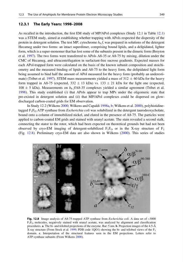
In Study 12.2 (Wilkens 2000; Wilkens and Capaldi 1998a, b; Wilkens et al. 2000), polyhistidinetagged F 1 F O ATP synthase from Escherichia coli was solubilized in the detergent taurodeoxycholate,
bound onto a column of immobilized nickel, and eluted in the presence of A8-75. The particles were
applied to carbon-coated EM grids and stained with uranyl acetate. The stain revealed a second stalk,
connecting the stator to the rotor, which had been expected on theoretical grounds but had not been
observed by cryo-EM imaging of detergent-solubilized F 1 F O or in the X-ray structure of F 1
(Fig. 12.8). Preliminary cryo-EM data are also shown in Wilkens (2000). This series of studies
Fig. 12.8 Image analysis of A8-75-trapped ATP synthase from Escherichia coli. A data set of ~3000
F 1 F O molecules, negatively stained with uranyl acetate, was analyzed by alignment and classification
procedures. a. The bi- and trilobed projections of the enzyme. Bar: 5 nm. b. Projection images of the 4.5-Å
X-ray structure (From Stock et al. 1999; PDB code 1QO1) showing the bi- and trilobed views of the F 1
domain. c. Interpretation of the structural features seen in the EM projections. Letters refer to
ATP-synthase subunits (From Wilkens 2000).
12.3 The Use of Amphipols for Membrane Protein Electron Microscopy Studies
549
As recalled in the introduction, the first EM study of MP/APol complexes (Study 12.1 in Table 12.1)
was a STEM study, aimed at establishing whether trapping with APols respected the dispersity of the
protein in detergent solution. The test MP, cytochrome b 6 f, was prepared in solutions of the detergent
Hecameg under two forms: an intact superdimer, comprising bound lipids, and a delipidated, lighter
form, which is a super-monomer that has lost some of the subunits present in the dimeric form (Breyton
et al. 1997). The two forms were transferred to APols A8-35 or A8-75 by mixing, dilution under the
CMC of Hecameg, and ultracentrifugation in surfactant-free sucrose gradients. Expected masses for
each APol-trapped form were calculated on the basis of the known subunit composition and stoichiometry and the measured binding of lipids and A8-75 to the heavy form, the delipidated light form
being assumed to bind half the amount of APol measured for the heavy form (probably an underestimate) (Tribet et al. 1997). STEM mass measurements yielded a mass of 312 Æ 60 kDa for the heavy
form trapped in A8-75 (expected, 332 Æ 13 kDa) vs. 133 Æ 21 kDa for the light one (expected,
108 Æ 5 kDa). Measurements on b 6 f/A8-35 complexes yielded a similar agreement (Tribet et al.
1998). This study established (i) that APols appear to trap MPs under the oligomeric state that
pre-existed in detergent solution and (ii) that MP/APol complexes could be dispersed on glowdischarged carbon-coated grids for EM observation.
In Study 12.2 (Wilkens 2000; Wilkens and Capaldi 1998a, b; Wilkens et al. 2000), polyhistidinetagged F 1 F O ATP synthase from Escherichia coli was solubilized in the detergent taurodeoxycholate,
bound onto a column of immobilized nickel, and eluted in the presence of A8-75. The particles were
applied to carbon-coated EM grids and stained with uranyl acetate. The stain revealed a second stalk,
connecting the stator to the rotor, which had been expected on theoretical grounds but had not been
observed by cryo-EM imaging of detergent-solubilized F 1 F O or in the X-ray structure of F 1
(Fig. 12.8). Preliminary cryo-EM data are also shown in Wilkens (2000). This series of studies
Fig. 12.8 Image analysis of A8-75-trapped ATP synthase from Escherichia coli. A data set of ~3000
F 1 F O molecules, negatively stained with uranyl acetate, was analyzed by alignment and classification
procedures. a. The bi- and trilobed projections of the enzyme. Bar: 5 nm. b. Projection images of the 4.5-Å
X-ray structure (From Stock et al. 1999; PDB code 1QO1) showing the bi- and trilobed views of the F 1
domain. c. Interpretation of the structural features seen in the EM projections. Letters refer to
ATP-synthase subunits (From Wilkens 2000).
12.3 The Use of Amphipols for Membrane Protein Electron Microscopy Studies
549