

surface, if not in the interior of MPs. The molecular details of protein/lipid interactions define their
binding sites, their specificity, and their modes of interaction. Many lipids, therefore, should be
considered as cofactors rather than a mere solvent. As for any ligand, lipids will stabilize preferentially
protein conformation(s) that provides them with the most affine binding sites. Thereby, they have the
potential both to stabilize MPs and to influence their conformational equilibria and, in so doing, to
modulate their function – a fact that has long been recognized by membrane biochemists (for a recent
example of allosteric regulation of a GPCR by cholesterol, see Casiraghi et al. 2016). As will be
discussed in Chap. 2, detergents compete with lipids for the hydrophobic TM surface of MPs. The
resulting delipidation is a major cause of the destabilization that is generally observed following
extraction of MPs from their native membrane environment using detergents.
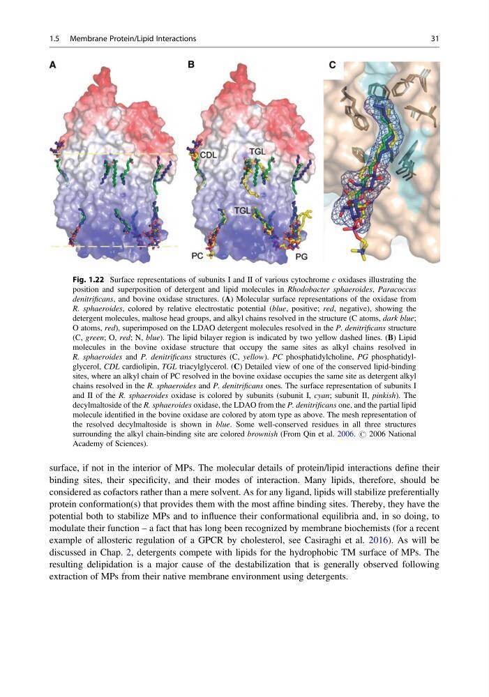
Fig. 1.22 Surface representations of subunits I and II of various cytochrome c oxidases illustrating the
position and superposition of detergent and lipid molecules in Rhodobacter sphaeroides, Paracoccus
denitrificans, and bovine oxidase structures. (A) Molecular surface representations of the oxidase from
R. sphaeroides, colored by relative electrostatic potential (blue, positive; red, negative), showing the
detergent molecules, maltose head groups, and alkyl chains resolved in the structure (C atoms, dark blue;
O atoms, red), superimposed on the LDAO detergent molecules resolved in the P. denitrificans structure
(C, green; O, red; N, blue). The lipid bilayer region is indicated by two yellow dashed lines. (B) Lipid
molecules in the bovine oxidase structure that occupy the same sites as alkyl chains resolved in
R. sphaeroides and P. denitrificans structures (C, yellow). PC phosphatidylcholine, PG phosphatidylglycerol, CDL cardiolipin, TGL triacylglycerol. (C) Detailed view of one of the conserved lipid-binding
sites, where an alkyl chain of PC resolved in the bovine oxidase occupies the same site as detergent alkyl
chains resolved in the R. sphaeroides and P. denitrificans ones. The surface representation of subunits I
and II of the R. sphaeroides oxidase is colored by subunits (subunit I, cyan; subunit II, pinkish). The
decylmaltoside of the R. sphaeroides oxidase, the LDAO from the P. denitrificans one, and the partial lipid
molecule identified in the bovine oxidase are colored by atom type as above. The mesh representation of
the resolved decylmaltoside is shown in blue. Some well-conserved residues in all three structures
surrounding the alkyl chain-binding site are colored brownish (From Qin et al. 2006. # 2006 National
Academy of Sciences).
1.5 Membrane Protein/Lipid Interactions
31
binding sites, their specificity, and their modes of interaction. Many lipids, therefore, should be
considered as cofactors rather than a mere solvent. As for any ligand, lipids will stabilize preferentially
protein conformation(s) that provides them with the most affine binding sites. Thereby, they have the
potential both to stabilize MPs and to influence their conformational equilibria and, in so doing, to
modulate their function – a fact that has long been recognized by membrane biochemists (for a recent
example of allosteric regulation of a GPCR by cholesterol, see Casiraghi et al. 2016). As will be
discussed in Chap. 2, detergents compete with lipids for the hydrophobic TM surface of MPs. The
resulting delipidation is a major cause of the destabilization that is generally observed following
extraction of MPs from their native membrane environment using detergents.
Fig. 1.22 Surface representations of subunits I and II of various cytochrome c oxidases illustrating the
position and superposition of detergent and lipid molecules in Rhodobacter sphaeroides, Paracoccus
denitrificans, and bovine oxidase structures. (A) Molecular surface representations of the oxidase from
R. sphaeroides, colored by relative electrostatic potential (blue, positive; red, negative), showing the
detergent molecules, maltose head groups, and alkyl chains resolved in the structure (C atoms, dark blue;
O atoms, red), superimposed on the LDAO detergent molecules resolved in the P. denitrificans structure
(C, green; O, red; N, blue). The lipid bilayer region is indicated by two yellow dashed lines. (B) Lipid
molecules in the bovine oxidase structure that occupy the same sites as alkyl chains resolved in
R. sphaeroides and P. denitrificans structures (C, yellow). PC phosphatidylcholine, PG phosphatidylglycerol, CDL cardiolipin, TGL triacylglycerol. (C) Detailed view of one of the conserved lipid-binding
sites, where an alkyl chain of PC resolved in the bovine oxidase occupies the same site as detergent alkyl
chains resolved in the R. sphaeroides and P. denitrificans ones. The surface representation of subunits I
and II of the R. sphaeroides oxidase is colored by subunits (subunit I, cyan; subunit II, pinkish). The
decylmaltoside of the R. sphaeroides oxidase, the LDAO from the P. denitrificans one, and the partial lipid
molecule identified in the bovine oxidase are colored by atom type as above. The mesh representation of
the resolved decylmaltoside is shown in blue. Some well-conserved residues in all three structures
surrounding the alkyl chain-binding site are colored brownish (From Qin et al. 2006. # 2006 National
Academy of Sciences).
1.5 Membrane Protein/Lipid Interactions
31