

is known to be required for the channel to be functional. In both the cytochrome bc 1 (Lange
et al. 2001) and cytochrome b 6 f (Kurisu et al. 2003; Stroebel et al. 2003) complexes, lipids
stabilize the assembly of the single TM helix of the Rieske iron-sulfur protein with the rest of
the complex. In some cases, e.g. subunit IV in the cytochrome c oxidase from Rhodobacter
sphaeroides (Svensson-Ek et al. 2002), interactions between a subunit and the rest of the
complex it is part of appear to be entirely mediated by lipids (see Palsdottir and Hunte 2004,
and references therein). As already mentioned, a continuous layer of lipids also seems to
separate the TM regions of Complex I, cytochrome bc 1 , and cytochrome c oxidase in the
mitochondrial respirasome (Althoff et al. 2011) (cf. Chap. 12, Fig. 12.17). Lipids similarly
mediate the assembly of BR trimers into 2D crystals in the purple membrane patches from
Halobacterium salinarum (§ 1.5.1).
• A less frequent but highly interesting case is that of lipids located inside a MP. The
arrangement of lipids in the 2.5-Å resolution crystal structure of Photosystem I reaction
center from Synechococcus elongatus offers three types of situation (Fig. 1.21). The reaction
center as a whole is a supertrimer (Fig. 1.21A), each supermonomer comprising 12 protein
subunits and no less than 127 cofactors: 96 chlorophylls, 2 phylloquinones, 3 Fe 4 S 4 clusters,
22 carotenoids, and 4 lipids. The four lipids, shown in turquoise in Fig. 1.21B, are three PG
molecules (numbered I, III, and IV) and a monogalactosyl diglyceride one (MGDG; numbered II). Lipid IV (a PG) occupies a rather classical location, being bound to the external,
membrane-exposed surface of the trimer. The other three lipids are buried, lipids I (a PG) and
II (the MGDG) inside each supermonomer and lipid III (a PG) at the interface between
supermonomers (Fig. 1.21B). Lipid III is particularly remarkable: as shown in Fig. 1.21C, it
does not simply fill a void between subunits; it acts as a bona fide cofactor, its phosphate
group providing the fifth ligand to the magnesium atom of one of the chlorophylls. Six more
lipids have been identified in the recent 2.8-Å structure of a PSI-LHCI supercomplex (Qin
et al. 2015). Other examples of protein-buried lipids are discussed in Palsdottir and
Hunte (2004).
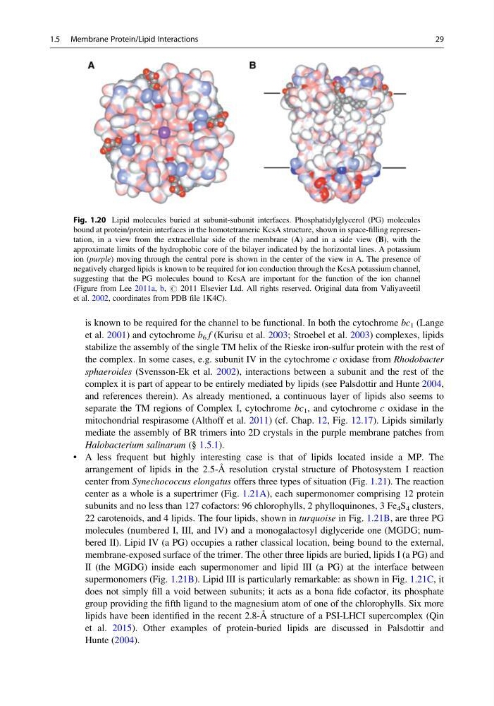
Fig. 1.20 Lipid molecules buried at subunit-subunit interfaces. Phosphatidylglycerol (PG) molecules
bound at protein/protein interfaces in the homotetrameric KcsA structure, shown in space-filling representation, in a view from the extracellular side of the membrane (A) and in a side view (B), with the
approximate limits of the hydrophobic core of the bilayer indicated by the horizontal lines. A potassium
ion (purple) moving through the central pore is shown in the center of the view in A. The presence of
negatively charged lipids is known to be required for ion conduction through the KcsA potassium channel,
suggesting that the PG molecules bound to KcsA are important for the function of the ion channel
(Figure from Lee 2011a, b, # 2011 Elsevier Ltd. All rights reserved. Original data from Valiyaveetil
et al. 2002, coordinates from PDB file 1K4C).
1.5 Membrane Protein/Lipid Interactions
29
et al. 2001) and cytochrome b 6 f (Kurisu et al. 2003; Stroebel et al. 2003) complexes, lipids
stabilize the assembly of the single TM helix of the Rieske iron-sulfur protein with the rest of
the complex. In some cases, e.g. subunit IV in the cytochrome c oxidase from Rhodobacter
sphaeroides (Svensson-Ek et al. 2002), interactions between a subunit and the rest of the
complex it is part of appear to be entirely mediated by lipids (see Palsdottir and Hunte 2004,
and references therein). As already mentioned, a continuous layer of lipids also seems to
separate the TM regions of Complex I, cytochrome bc 1 , and cytochrome c oxidase in the
mitochondrial respirasome (Althoff et al. 2011) (cf. Chap. 12, Fig. 12.17). Lipids similarly
mediate the assembly of BR trimers into 2D crystals in the purple membrane patches from
Halobacterium salinarum (§ 1.5.1).
• A less frequent but highly interesting case is that of lipids located inside a MP. The
arrangement of lipids in the 2.5-Å resolution crystal structure of Photosystem I reaction
center from Synechococcus elongatus offers three types of situation (Fig. 1.21). The reaction
center as a whole is a supertrimer (Fig. 1.21A), each supermonomer comprising 12 protein
subunits and no less than 127 cofactors: 96 chlorophylls, 2 phylloquinones, 3 Fe 4 S 4 clusters,
22 carotenoids, and 4 lipids. The four lipids, shown in turquoise in Fig. 1.21B, are three PG
molecules (numbered I, III, and IV) and a monogalactosyl diglyceride one (MGDG; numbered II). Lipid IV (a PG) occupies a rather classical location, being bound to the external,
membrane-exposed surface of the trimer. The other three lipids are buried, lipids I (a PG) and
II (the MGDG) inside each supermonomer and lipid III (a PG) at the interface between
supermonomers (Fig. 1.21B). Lipid III is particularly remarkable: as shown in Fig. 1.21C, it
does not simply fill a void between subunits; it acts as a bona fide cofactor, its phosphate
group providing the fifth ligand to the magnesium atom of one of the chlorophylls. Six more
lipids have been identified in the recent 2.8-Å structure of a PSI-LHCI supercomplex (Qin
et al. 2015). Other examples of protein-buried lipids are discussed in Palsdottir and
Hunte (2004).
Fig. 1.20 Lipid molecules buried at subunit-subunit interfaces. Phosphatidylglycerol (PG) molecules
bound at protein/protein interfaces in the homotetrameric KcsA structure, shown in space-filling representation, in a view from the extracellular side of the membrane (A) and in a side view (B), with the
approximate limits of the hydrophobic core of the bilayer indicated by the horizontal lines. A potassium
ion (purple) moving through the central pore is shown in the center of the view in A. The presence of
negatively charged lipids is known to be required for ion conduction through the KcsA potassium channel,
suggesting that the PG molecules bound to KcsA are important for the function of the ion channel
(Figure from Lee 2011a, b, # 2011 Elsevier Ltd. All rights reserved. Original data from Valiyaveetil
et al. 2002, coordinates from PDB file 1K4C).
1.5 Membrane Protein/Lipid Interactions
29