

briefly discuss below, and factors linked to crystallography itself, such as the development of
crystallization robots, of ever more powerful synchrotrons with ever brighter and finer beams, and,
along with it, the ability to prepare, recover, identify, select, and analyze smaller and smaller crystals in
a more and more automated manner. Technological progress in these directions is constant, with the
use of micrometric beams and crystals becoming almost routine.
The most recent major technical progress, currently at its onset and in full development, is the
use of free-electron laser X-ray (X-FEL) sources, which deliver very short (~50 to 100 fs) pulses whose
brightness – many orders of magnitude above that of synchrotron sources – makes it possible to record
fragmentary diffraction patterns from thousands if not millions of micrometric to submicrometric
crystals and, by merging them, to reconstruct the full pattern and calculate the electron density map of
the target protein. Data are collected in the first tens of femtoseconds of exposure, i.e. before radiation
damage sets in (see Neutze et al. 2000, 2015; Chapman et al. 2011; Boutet et al. 2012; Johansson et al.
2012; Kang et al. 2013; Martin-Garcia et al. 2016). Despite the limited number of X-FEL sources, their
high running costs, and the considerable technical difficulties involved, this approach, which has been
proven to be applicable to MPs (Chapman et al. 2011; Johansson et al. 2012; Kupitz et al. 2014; Suga
et al. 2017), has the potential of yielding the structures of important MP targets that are reluctant to
form crystals large enough for conventional analysis using synchrotron X-ray sources.
Among biochemical progresses, a decisive one was the ability to produce, by overexpression,
MPs that are too rare in natural organisms to be purified in amounts commensurate to the needs of a
crystallographic study (see Chap. 1), combined with the miniaturization and robotization of the
crystallization assays, which multiplies the number of trials that can be conducted with a given amount
of protein. Another useful progress has been the diversification of surfactants (reviewed in Zhang et al.
2011; Sadaf et al. 2015; cf. Chap. 2, § 2.5.1), even though, compared to the tens of novel molecules
that have been designed and validated biochemically, their contribution to the determination of novel
MP structures remains, to date, relatively modest (see Parker and Newstead 2012; Loll 2014; Moraes
et al. 2014). An example of success story is the use of “MNG” (maltose-neopentyl glycol) detergents
(see Chap. 2, Fig. 2.15) for handling and crystallizing G protein-coupled receptors (GPCRs)
(Rasmussen et al. 2007, 2011; Chae et al. 2010). A highly significant progress has been the development of nonconventional approaches to crystallization. Because those bear more directly on the
0
0
100
200
300
400
500
600
700
5
1 0
1 5
expected exponential growth
at year 20 (2005)
Stephen White Lab at UC Irvine
http://blanco.biomol.uci.edu/mpstruc
years since first structure [1985]
2016
n = 672
cumulative unique MP structures (n)
20
30
25
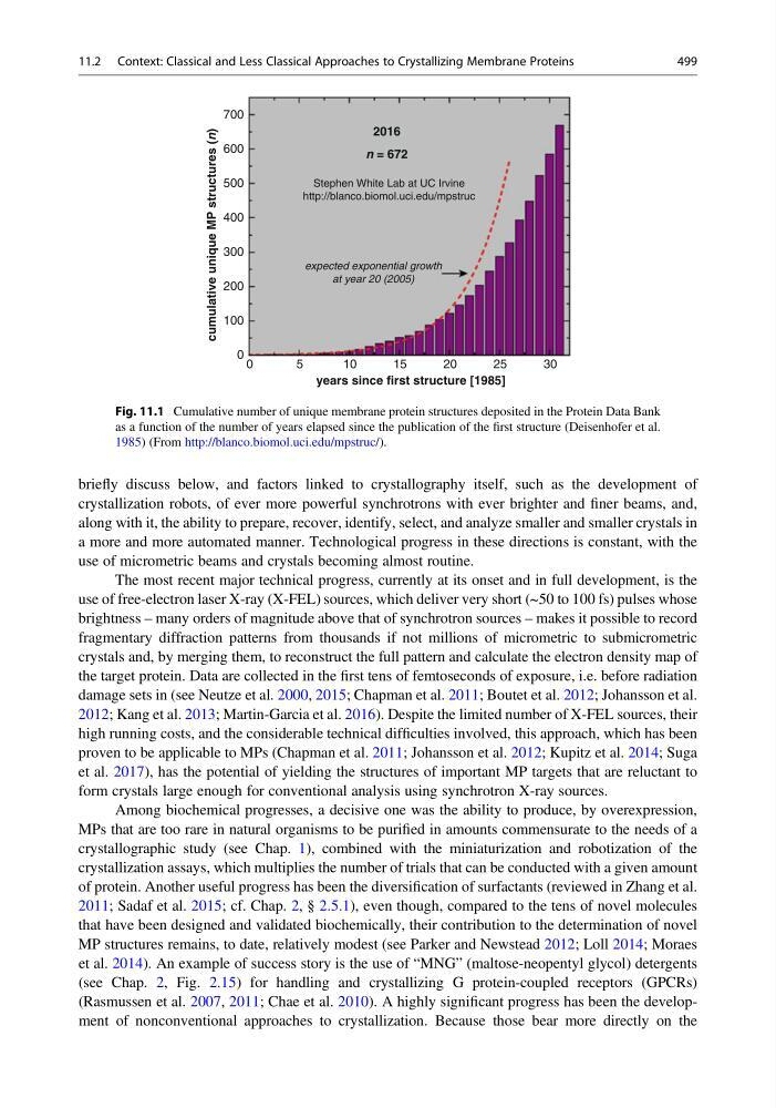
Fig. 11.1 Cumulative number of unique membrane protein structures deposited in the Protein Data Bank
as a function of the number of years elapsed since the publication of the first structure (Deisenhofer et al.
1985) (From http://blanco.biomol.uci.edu/mpstruc/).
11.2 Context: Classical and Less Classical Approaches to Crystallizing Membrane Proteins
499
crystallization robots, of ever more powerful synchrotrons with ever brighter and finer beams, and,
along with it, the ability to prepare, recover, identify, select, and analyze smaller and smaller crystals in
a more and more automated manner. Technological progress in these directions is constant, with the
use of micrometric beams and crystals becoming almost routine.
The most recent major technical progress, currently at its onset and in full development, is the
use of free-electron laser X-ray (X-FEL) sources, which deliver very short (~50 to 100 fs) pulses whose
brightness – many orders of magnitude above that of synchrotron sources – makes it possible to record
fragmentary diffraction patterns from thousands if not millions of micrometric to submicrometric
crystals and, by merging them, to reconstruct the full pattern and calculate the electron density map of
the target protein. Data are collected in the first tens of femtoseconds of exposure, i.e. before radiation
damage sets in (see Neutze et al. 2000, 2015; Chapman et al. 2011; Boutet et al. 2012; Johansson et al.
2012; Kang et al. 2013; Martin-Garcia et al. 2016). Despite the limited number of X-FEL sources, their
high running costs, and the considerable technical difficulties involved, this approach, which has been
proven to be applicable to MPs (Chapman et al. 2011; Johansson et al. 2012; Kupitz et al. 2014; Suga
et al. 2017), has the potential of yielding the structures of important MP targets that are reluctant to
form crystals large enough for conventional analysis using synchrotron X-ray sources.
Among biochemical progresses, a decisive one was the ability to produce, by overexpression,
MPs that are too rare in natural organisms to be purified in amounts commensurate to the needs of a
crystallographic study (see Chap. 1), combined with the miniaturization and robotization of the
crystallization assays, which multiplies the number of trials that can be conducted with a given amount
of protein. Another useful progress has been the diversification of surfactants (reviewed in Zhang et al.
2011; Sadaf et al. 2015; cf. Chap. 2, § 2.5.1), even though, compared to the tens of novel molecules
that have been designed and validated biochemically, their contribution to the determination of novel
MP structures remains, to date, relatively modest (see Parker and Newstead 2012; Loll 2014; Moraes
et al. 2014). An example of success story is the use of “MNG” (maltose-neopentyl glycol) detergents
(see Chap. 2, Fig. 2.15) for handling and crystallizing G protein-coupled receptors (GPCRs)
(Rasmussen et al. 2007, 2011; Chae et al. 2010). A highly significant progress has been the development of nonconventional approaches to crystallization. Because those bear more directly on the
0
0
100
200
300
400
500
600
700
5
1 0
1 5
expected exponential growth
at year 20 (2005)
Stephen White Lab at UC Irvine
http://blanco.biomol.uci.edu/mpstruc
years since first structure [1985]
2016
n = 672
cumulative unique MP structures (n)
20
30
25
Fig. 11.1 Cumulative number of unique membrane protein structures deposited in the Protein Data Bank
as a function of the number of years elapsed since the publication of the first structure (Deisenhofer et al.
1985) (From http://blanco.biomol.uci.edu/mpstruc/).
11.2 Context: Classical and Less Classical Approaches to Crystallizing Membrane Proteins
499