

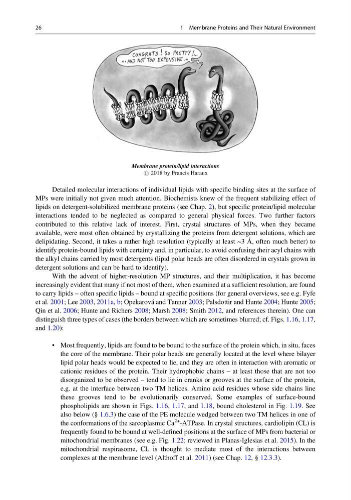
Membrane protein/lipid interactions
# 2018 by Francis Haraux
Detailed molecular interactions of individual lipids with specific binding sites at the surface of
MPs were initially not given much attention. Biochemists knew of the frequent stabilizing effect of
lipids on detergent-solubilized membrane proteins (see Chap. 2), but specific protein/lipid molecular
interactions tended to be neglected as compared to general physical forces. Two further factors
contributed to this relative lack of interest. First, crystal structures of MPs, when they became
available, were most often obtained by crystallizing the proteins from detergent solutions, which are
delipidating. Second, it takes a rather high resolution (typically at least ~3 Å, often much better) to
identify protein-bound lipids with certainty and, in particular, to avoid confusing their acyl chains with
the alkyl chains carried by most detergents (lipid polar heads are often disordered in crystals grown in
detergent solutions and can be hard to identify).
With the advent of higher-resolution MP structures, and their multiplication, it has become
increasingly evident that many if not most of them, when examined at a sufficient resolution, are found
to carry lipids – often specific lipids – bound at specific positions (for general overviews, see e.g. Fyfe
et al. 2001; Lee 2003, 2011a, b; Opekarová and Tanner 2003; Palsdottir and Hunte 2004; Hunte 2005;
Qin et al. 2006; Hunte and Richers 2008; Marsh 2008; Smith 2012, and references therein). One can
distinguish three types of cases (the borders between which are sometimes blurred; cf. Figs. 1.16, 1.17,
and 1.20):
• Most frequently, lipids are found to be bound to the surface of the protein which, in situ, faces
the core of the membrane. Their polar heads are generally located at the level where bilayer
lipid polar heads would be expected to lie, and they are often in interaction with aromatic or
cationic residues of the protein. Their hydrophobic chains – at least those that are not too
disorganized to be observed – tend to lie in cranks or grooves at the surface of the protein,
e.g. at the interface between two TM helices. Amino acid residues whose side chains line
these grooves tend to be evolutionarily conserved. Some examples of surface-bound
phospholipids are shown in Figs. 1.16, 1.17, and 1.18, bound cholesterol in Fig. 1.19. See
also below (§ 1.6.3) the case of the PE molecule wedged between two TM helices in one of
the conformations of the sarcoplasmic Ca
2+ -ATPase. In crystal structures, cardiolipin (CL) is
frequently found to be bound at well-defined positions at the surface of MPs from bacterial or
mitochondrial membranes (see e.g. Fig. 1.22; reviewed in Planas-Iglesias et al. 2015). In the
mitochondrial respirasome, CL is thought to mediate most of the interactions between
complexes at the membrane level (Althoff et al. 2011) (see Chap. 12, § 12.3.3).
26
1 Membrane Proteins and Their Natural Environment
# 2018 by Francis Haraux
Detailed molecular interactions of individual lipids with specific binding sites at the surface of
MPs were initially not given much attention. Biochemists knew of the frequent stabilizing effect of
lipids on detergent-solubilized membrane proteins (see Chap. 2), but specific protein/lipid molecular
interactions tended to be neglected as compared to general physical forces. Two further factors
contributed to this relative lack of interest. First, crystal structures of MPs, when they became
available, were most often obtained by crystallizing the proteins from detergent solutions, which are
delipidating. Second, it takes a rather high resolution (typically at least ~3 Å, often much better) to
identify protein-bound lipids with certainty and, in particular, to avoid confusing their acyl chains with
the alkyl chains carried by most detergents (lipid polar heads are often disordered in crystals grown in
detergent solutions and can be hard to identify).
With the advent of higher-resolution MP structures, and their multiplication, it has become
increasingly evident that many if not most of them, when examined at a sufficient resolution, are found
to carry lipids – often specific lipids – bound at specific positions (for general overviews, see e.g. Fyfe
et al. 2001; Lee 2003, 2011a, b; Opekarová and Tanner 2003; Palsdottir and Hunte 2004; Hunte 2005;
Qin et al. 2006; Hunte and Richers 2008; Marsh 2008; Smith 2012, and references therein). One can
distinguish three types of cases (the borders between which are sometimes blurred; cf. Figs. 1.16, 1.17,
and 1.20):
• Most frequently, lipids are found to be bound to the surface of the protein which, in situ, faces
the core of the membrane. Their polar heads are generally located at the level where bilayer
lipid polar heads would be expected to lie, and they are often in interaction with aromatic or
cationic residues of the protein. Their hydrophobic chains – at least those that are not too
disorganized to be observed – tend to lie in cranks or grooves at the surface of the protein,
e.g. at the interface between two TM helices. Amino acid residues whose side chains line
these grooves tend to be evolutionarily conserved. Some examples of surface-bound
phospholipids are shown in Figs. 1.16, 1.17, and 1.18, bound cholesterol in Fig. 1.19. See
also below (§ 1.6.3) the case of the PE molecule wedged between two TM helices in one of
the conformations of the sarcoplasmic Ca
2+ -ATPase. In crystal structures, cardiolipin (CL) is
frequently found to be bound at well-defined positions at the surface of MPs from bacterial or
mitochondrial membranes (see e.g. Fig. 1.22; reviewed in Planas-Iglesias et al. 2015). In the
mitochondrial respirasome, CL is thought to mediate most of the interactions between
complexes at the membrane level (Althoff et al. 2011) (see Chap. 12, § 12.3.3).
26
1 Membrane Proteins and Their Natural Environment