

There are two basic types of NMR studies, solution NMR (sNMR) and “solid-state” NMR
(ssNMR) studies, the latter actually applying both to solid, e.g. frozen, samples and to viscous ones,
such as a biological membrane at room temperature, as well as to large macromolecules that are
beyond the reach of sNMR. A major difference between the two approaches lies in the way the
influence of the magnetic field of the spectrometer is averaged over the various orientations the target
macromolecule can adopt with respect to it. This will define the average field experienced by each
nucleus, and, thereby, the frequency at which it resonates when subjected to an oscillating field.
Solution NMR relies on the fast tumbling of the molecule, which must be rapid enough so that, on
the time-scale of spin relaxation, each nucleus has experienced and averaged the influence of all
possible orientations in the magnetic field. If this condition is not met, various equivalent nuclei in
various molecules will experience a slightly different average field and resonate at slightly different
frequencies, resulting in peak broadening. Because there are tens if not hundreds of similar nuclei,
e.g. amide protons, in even a small protein, and many of them resonate at frequencies that are not spread
over a very broad range, peak broadening prevents separating the signals from various nuclei from one
another and identifying them, which is a prerequisite to any structure determination. In sNMR,
averaging is left to the molecule itself, which, by rapidly tumbling, experiences all possible orientations.
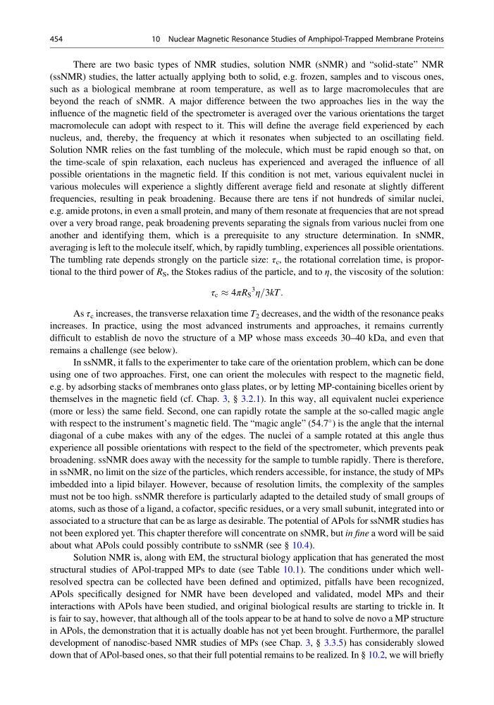
The tumbling rate depends strongly on the particle size: τ c , the rotational correlation time, is proportional to the third power of R S , the Stokes radius of the particle, and to η, the viscosity of the solution:
τ c % 4πR S
3
η=3kT:
As τ c increases, the transverse relaxation time T 2 decreases, and the width of the resonance peaks
increases. In practice, using the most advanced instruments and approaches, it remains currently
difficult to establish de novo the structure of a MP whose mass exceeds 30–40 kDa, and even that
remains a challenge (see below).
In ssNMR, it falls to the experimenter to take care of the orientation problem, which can be done
using one of two approaches. First, one can orient the molecules with respect to the magnetic field,
e.g. by adsorbing stacks of membranes onto glass plates, or by letting MP-containing bicelles orient by
themselves in the magnetic field (cf. Chap. 3, § 3.2.1). In this way, all equivalent nuclei experience
(more or less) the same field. Second, one can rapidly rotate the sample at the so-called magic angle
with respect to the instrument’s magnetic field. The “magic angle” (54.7
) is the angle that the internal
diagonal of a cube makes with any of the edges. The nuclei of a sample rotated at this angle thus
experience all possible orientations with respect to the field of the spectrometer, which prevents peak
broadening. ssNMR does away with the necessity for the sample to tumble rapidly. There is therefore,
in ssNMR, no limit on the size of the particles, which renders accessible, for instance, the study of MPs
imbedded into a lipid bilayer. However, because of resolution limits, the complexity of the samples
must not be too high. ssNMR therefore is particularly adapted to the detailed study of small groups of
atoms, such as those of a ligand, a cofactor, specific residues, or a very small subunit, integrated into or
associated to a structure that can be as large as desirable. The potential of APols for ssNMR studies has
not been explored yet. This chapter therefore will concentrate on sNMR, but in fine a word will be said
about what APols could possibly contribute to ssNMR (see § 10.4).
Solution NMR is, along with EM, the structural biology application that has generated the most
structural studies of APol-trapped MPs to date (see Table 10.1). The conditions under which wellresolved spectra can be collected have been defined and optimized, pitfalls have been recognized,
APols specifically designed for NMR have been developed and validated, model MPs and their
interactions with APols have been studied, and original biological results are starting to trickle in. It
is fair to say, however, that although all of the tools appear to be at hand to solve de novo a MP structure
in APols, the demonstration that it is actually doable has not yet been brought. Furthermore, the parallel
development of nanodisc-based NMR studies of MPs (see Chap. 3, § 3.3.5) has considerably slowed
down that of APol-based ones, so that their full potential remains to be realized. In § 10.2, we will briefly
454
10 Nuclear Magnetic Resonance Studies of Amphipol-Trapped Membrane Proteins
(ssNMR) studies, the latter actually applying both to solid, e.g. frozen, samples and to viscous ones,
such as a biological membrane at room temperature, as well as to large macromolecules that are
beyond the reach of sNMR. A major difference between the two approaches lies in the way the
influence of the magnetic field of the spectrometer is averaged over the various orientations the target
macromolecule can adopt with respect to it. This will define the average field experienced by each
nucleus, and, thereby, the frequency at which it resonates when subjected to an oscillating field.
Solution NMR relies on the fast tumbling of the molecule, which must be rapid enough so that, on
the time-scale of spin relaxation, each nucleus has experienced and averaged the influence of all
possible orientations in the magnetic field. If this condition is not met, various equivalent nuclei in
various molecules will experience a slightly different average field and resonate at slightly different
frequencies, resulting in peak broadening. Because there are tens if not hundreds of similar nuclei,
e.g. amide protons, in even a small protein, and many of them resonate at frequencies that are not spread
over a very broad range, peak broadening prevents separating the signals from various nuclei from one
another and identifying them, which is a prerequisite to any structure determination. In sNMR,
averaging is left to the molecule itself, which, by rapidly tumbling, experiences all possible orientations.
The tumbling rate depends strongly on the particle size: τ c , the rotational correlation time, is proportional to the third power of R S , the Stokes radius of the particle, and to η, the viscosity of the solution:
τ c % 4πR S
3
η=3kT:
As τ c increases, the transverse relaxation time T 2 decreases, and the width of the resonance peaks
increases. In practice, using the most advanced instruments and approaches, it remains currently
difficult to establish de novo the structure of a MP whose mass exceeds 30–40 kDa, and even that
remains a challenge (see below).
In ssNMR, it falls to the experimenter to take care of the orientation problem, which can be done
using one of two approaches. First, one can orient the molecules with respect to the magnetic field,
e.g. by adsorbing stacks of membranes onto glass plates, or by letting MP-containing bicelles orient by
themselves in the magnetic field (cf. Chap. 3, § 3.2.1). In this way, all equivalent nuclei experience
(more or less) the same field. Second, one can rapidly rotate the sample at the so-called magic angle
with respect to the instrument’s magnetic field. The “magic angle” (54.7
) is the angle that the internal
diagonal of a cube makes with any of the edges. The nuclei of a sample rotated at this angle thus
experience all possible orientations with respect to the field of the spectrometer, which prevents peak
broadening. ssNMR does away with the necessity for the sample to tumble rapidly. There is therefore,
in ssNMR, no limit on the size of the particles, which renders accessible, for instance, the study of MPs
imbedded into a lipid bilayer. However, because of resolution limits, the complexity of the samples
must not be too high. ssNMR therefore is particularly adapted to the detailed study of small groups of
atoms, such as those of a ligand, a cofactor, specific residues, or a very small subunit, integrated into or
associated to a structure that can be as large as desirable. The potential of APols for ssNMR studies has
not been explored yet. This chapter therefore will concentrate on sNMR, but in fine a word will be said
about what APols could possibly contribute to ssNMR (see § 10.4).
Solution NMR is, along with EM, the structural biology application that has generated the most
structural studies of APol-trapped MPs to date (see Table 10.1). The conditions under which wellresolved spectra can be collected have been defined and optimized, pitfalls have been recognized,
APols specifically designed for NMR have been developed and validated, model MPs and their
interactions with APols have been studied, and original biological results are starting to trickle in. It
is fair to say, however, that although all of the tools appear to be at hand to solve de novo a MP structure
in APols, the demonstration that it is actually doable has not yet been brought. Furthermore, the parallel
development of nanodisc-based NMR studies of MPs (see Chap. 3, § 3.3.5) has considerably slowed
down that of APol-based ones, so that their full potential remains to be realized. In § 10.2, we will briefly
454
10 Nuclear Magnetic Resonance Studies of Amphipol-Trapped Membrane Proteins