

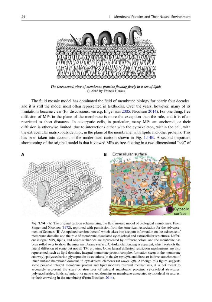
The (erroneous) view of membrane proteins floating freely in a sea of lipids
# 2018 by Francis Haraux
The fluid mosaic model has dominated the field of membrane biology for nearly four decades,
and it is still the model most often represented in textbooks. Over the years, however, many of its
limitations became clear (for discussions, see e.g. Engelman 2005; Nicolson 2014). For one thing, free
diffusion of MPs in the plane of the membrane is more the exception than the rule, and it is often
restricted to short distances. In eukaryotic cells, in particular, many MPs are anchored, or their
diffusion is otherwise limited, due to interactions either with the cytoskeleton, within the cell, with
the extracellular matrix, outside it, or, in the plane of the membrane, with lipids and other proteins. This
has been taken into account in the modernized cartoon shown in Fig. 1.14B. A second important
shortcoming of the original model is that it viewed MPs as free-floating in a two-dimensional “sea” of
Fig. 1.14 (A) The original cartoon schematizing the fluid mosaic model of biological membranes. From
Singer and Nicolson (1972), reprinted with permission from the American Association for the Advancement of Science. (B) An updated version thereof, which takes into account information on the existence of
membrane domains and the role of membrane-associated cytoskeletal and extracellular structures. Different integral MPs, lipids, and oligosaccharides are represented by different colors, and the membrane has
been rolled over to show the inner membrane surface. Cytoskeletal fencing is apparent, which restricts the
lateral diffusion of some but not all TM proteins. Other lateral diffusion restriction mechanisms are also
represented, such as lipid domains, integral membrane protein complex formation (seen in the membrane
cutaway), polysaccharide-glycoprotein associations (at the far top left), and direct or indirect attachment of
inner surface membrane domains to cytoskeletal elements (at lower left). Although this figure suggests
some possible integral membrane protein and lipid mobility restraint mechanisms, it is not meant to
accurately represent the sizes or structures of integral membrane proteins, cytoskeletal structures,
polysaccharides, lipids, submicro- or nano-sized domains or membrane-associated cytoskeletal structures,
or their crowding in the membrane (From Nicolson 2014).
24
1 Membrane Proteins and Their Natural Environment
# 2018 by Francis Haraux
The fluid mosaic model has dominated the field of membrane biology for nearly four decades,
and it is still the model most often represented in textbooks. Over the years, however, many of its
limitations became clear (for discussions, see e.g. Engelman 2005; Nicolson 2014). For one thing, free
diffusion of MPs in the plane of the membrane is more the exception than the rule, and it is often
restricted to short distances. In eukaryotic cells, in particular, many MPs are anchored, or their
diffusion is otherwise limited, due to interactions either with the cytoskeleton, within the cell, with
the extracellular matrix, outside it, or, in the plane of the membrane, with lipids and other proteins. This
has been taken into account in the modernized cartoon shown in Fig. 1.14B. A second important
shortcoming of the original model is that it viewed MPs as free-floating in a two-dimensional “sea” of
Fig. 1.14 (A) The original cartoon schematizing the fluid mosaic model of biological membranes. From
Singer and Nicolson (1972), reprinted with permission from the American Association for the Advancement of Science. (B) An updated version thereof, which takes into account information on the existence of
membrane domains and the role of membrane-associated cytoskeletal and extracellular structures. Different integral MPs, lipids, and oligosaccharides are represented by different colors, and the membrane has
been rolled over to show the inner membrane surface. Cytoskeletal fencing is apparent, which restricts the
lateral diffusion of some but not all TM proteins. Other lateral diffusion restriction mechanisms are also
represented, such as lipid domains, integral membrane protein complex formation (seen in the membrane
cutaway), polysaccharide-glycoprotein associations (at the far top left), and direct or indirect attachment of
inner surface membrane domains to cytoskeletal elements (at lower left). Although this figure suggests
some possible integral membrane protein and lipid mobility restraint mechanisms, it is not meant to
accurately represent the sizes or structures of integral membrane proteins, cytoskeletal structures,
polysaccharides, lipids, submicro- or nano-sized domains or membrane-associated cytoskeletal structures,
or their crowding in the membrane (From Nicolson 2014).
24
1 Membrane Proteins and Their Natural Environment