

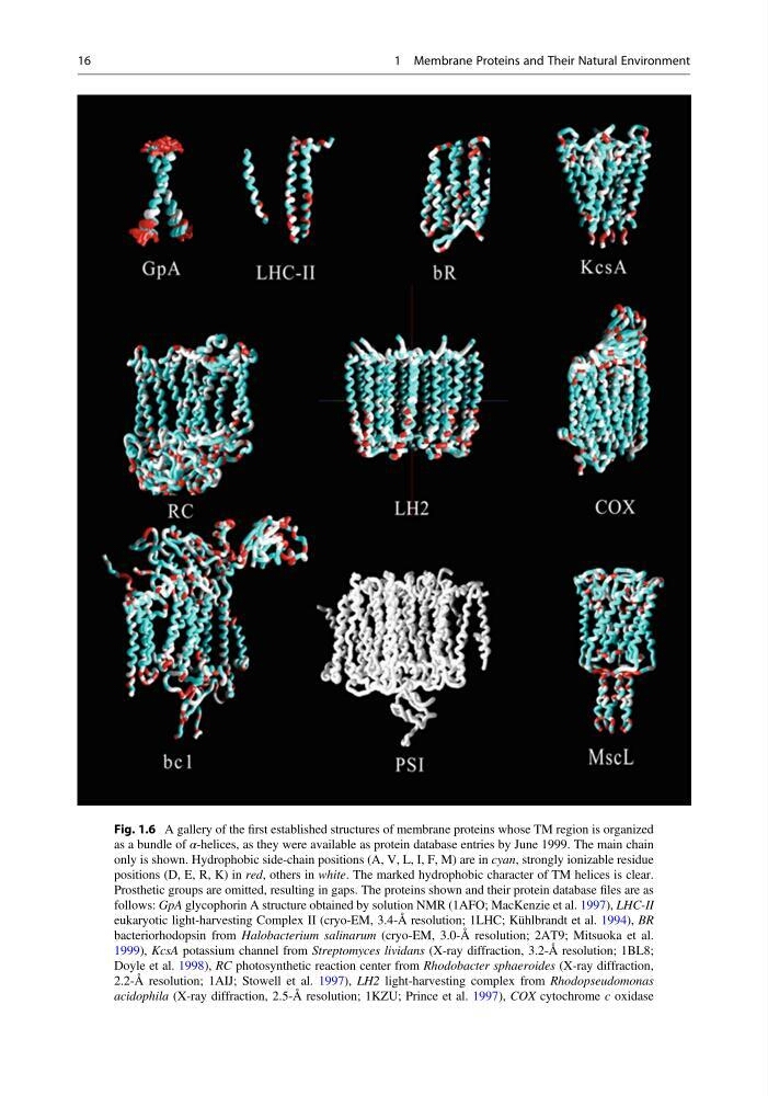
Fig. 1.6 A gallery of the first established structures of membrane proteins whose TM region is organized
as a bundle of α-helices, as they were available as protein database entries by June 1999. The main chain
only is shown. Hydrophobic side-chain positions (A, V, L, I, F, M) are in cyan, strongly ionizable residue
positions (D, E, R, K) in red, others in white. The marked hydrophobic character of TM helices is clear.
Prosthetic groups are omitted, resulting in gaps. The proteins shown and their protein database files are as
follows: GpA glycophorin A structure obtained by solution NMR (1AFO; MacKenzie et al. 1997), LHC-II
eukaryotic light-harvesting Complex II (cryo-EM, 3.4-Å resolution; 1LHC; Kühlbrandt et al. 1994), BR
bacteriorhodopsin from Halobacterium salinarum (cryo-EM, 3.0-Å resolution; 2AT9; Mitsuoka et al.
1999), KcsA potassium channel from Streptomyces lividans (X-ray diffraction, 3.2-Å resolution; 1BL8;
Doyle et al. 1998), RC photosynthetic reaction center from Rhodobacter sphaeroides (X-ray diffraction,
2.2-Å resolution; 1AIJ; Stowell et al. 1997), LH2 light-harvesting complex from Rhodopseudomonas
acidophila (X-ray diffraction, 2.5-Å resolution; 1KZU; Prince et al. 1997), COX cytochrome c oxidase
16
1 Membrane Proteins and Their Natural Environment
as a bundle of α-helices, as they were available as protein database entries by June 1999. The main chain
only is shown. Hydrophobic side-chain positions (A, V, L, I, F, M) are in cyan, strongly ionizable residue
positions (D, E, R, K) in red, others in white. The marked hydrophobic character of TM helices is clear.
Prosthetic groups are omitted, resulting in gaps. The proteins shown and their protein database files are as
follows: GpA glycophorin A structure obtained by solution NMR (1AFO; MacKenzie et al. 1997), LHC-II
eukaryotic light-harvesting Complex II (cryo-EM, 3.4-Å resolution; 1LHC; Kühlbrandt et al. 1994), BR
bacteriorhodopsin from Halobacterium salinarum (cryo-EM, 3.0-Å resolution; 2AT9; Mitsuoka et al.
1999), KcsA potassium channel from Streptomyces lividans (X-ray diffraction, 3.2-Å resolution; 1BL8;
Doyle et al. 1998), RC photosynthetic reaction center from Rhodobacter sphaeroides (X-ray diffraction,
2.2-Å resolution; 1AIJ; Stowell et al. 1997), LH2 light-harvesting complex from Rhodopseudomonas
acidophila (X-ray diffraction, 2.5-Å resolution; 1KZU; Prince et al. 1997), COX cytochrome c oxidase
16
1 Membrane Proteins and Their Natural Environment