

6.3.1.2.3 Kinetics and Thermodynamics of Folding OmpA in A8-35
A detailed study of the kinetics and thermodynamics of folding and unfolding of OmpA in A8-35 has
been carried out by Jörg H. Kleinschmidt and coworkers (Pocanschi et al. 2013). Folding was initiated
by 18-fold dilution of urea in the presence of various mass ratios of A8-35, ranging from 0.5 to 16 g per
g of OmpA and at various temperatures. Analyses of the time course of electrophoretic mobility shift
from the unfolded form (M r % 35 kDa) to the folded one (M r % 30 kDa) indicated that folding takes ~6
to 8 h at pH 10 (a high pH used to ensure a good solubility of the unfolded protein). The minimum mass
ratio of A8-35/OmpA required to achieve complete folding was 2:1. Kinetics of folding were obtained
by fluorescence spectroscopy and by monitoring the formation of tertiary structure by SDS-PAGE.
Whichever method was used, two parallel pathways of folding were observed, presumably due to the
coexistence, at the time of urea dilution, of various forms of OmpA carrying different charges and, as a
consequence, engaging in different folding paths. The rate constants of the two folding processes did
not depend on the concentration of A8-35, indicating that intermolecular interactions between proteins
are not involved. SDS-PAGE did not reveal any folding intermediates. (Such intermediates likely
exist, but they do not survive exposure to SDS and revert to the unfolded form.)
The activation energy of folding was determined from the temperature dependence of the folding
kinetics. The SDS-PAGE assay yielded an activation energy of ~5.9 Æ 4.1 kJÁmol
À1 (~1.4 kcalÁmol
À1
)
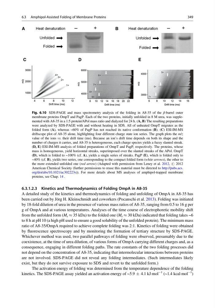
Fig. 6.10 SDS-PAGE and mass spectrometry analysis of the folding in A8-35 of the β-barrel outer
membrane proteins OmpT and PagP. Each of the two proteins, initially unfolded in 8 M urea, was supplemented with A8-35 in a 1:5 protein/APol mass ratio and dialyzed for 24 h. (A, B) The resulting preparations
were analyzed by SDS-PAGE with and without heating in SDS. All of unheated OmpT migrates as the
folded form (A), whereas ~60% of PagP has not reached its native conformation (B). (C) ESI-IM-MS
driftscope plot of A8-35 alone, highlighting four different charge state ion series. The graph plots the m/z
value of the ions vs. their drift time (ms). Because an ion’s drift time depends on both its shape and the
number of charges it carries, and A8-35 is heterogeneous, each charge species yields a fuzzy slanted streak.
(D, E) ESI-IM-MS analysis of folded preparations of OmpT and PagP, respectively. The proteins, whose
mass is homogeneous, yield horizontal streaks, superimposed over the slanted streaks of the APol. OmpT
(D), which is folded to ~100% (cf. A), yields a single series of streaks. PagP (E), which is folded only to
~40% (cf. B), yields two series, one corresponding to the compact folded form (white arrows), the other to
the more extended unfolded one (red arrow) (Adapted with permission from Leney et al. 2012, # 2012
American Chemical Society (further permissions to reuse this material must be directed to http://pubs.acs.
org/doi/abs/10.1021/ac302223s)). For more details about MS analyses of amphipol-trapped membrane
proteins, see Chap. 14 .
6.3 Amphipol-Assisted Folding of Membrane Proteins
349
A detailed study of the kinetics and thermodynamics of folding and unfolding of OmpA in A8-35 has
been carried out by Jörg H. Kleinschmidt and coworkers (Pocanschi et al. 2013). Folding was initiated
by 18-fold dilution of urea in the presence of various mass ratios of A8-35, ranging from 0.5 to 16 g per
g of OmpA and at various temperatures. Analyses of the time course of electrophoretic mobility shift
from the unfolded form (M r % 35 kDa) to the folded one (M r % 30 kDa) indicated that folding takes ~6
to 8 h at pH 10 (a high pH used to ensure a good solubility of the unfolded protein). The minimum mass
ratio of A8-35/OmpA required to achieve complete folding was 2:1. Kinetics of folding were obtained
by fluorescence spectroscopy and by monitoring the formation of tertiary structure by SDS-PAGE.
Whichever method was used, two parallel pathways of folding were observed, presumably due to the
coexistence, at the time of urea dilution, of various forms of OmpA carrying different charges and, as a
consequence, engaging in different folding paths. The rate constants of the two folding processes did
not depend on the concentration of A8-35, indicating that intermolecular interactions between proteins
are not involved. SDS-PAGE did not reveal any folding intermediates. (Such intermediates likely
exist, but they do not survive exposure to SDS and revert to the unfolded form.)
The activation energy of folding was determined from the temperature dependence of the folding
kinetics. The SDS-PAGE assay yielded an activation energy of ~5.9 Æ 4.1 kJÁmol
À1 (~1.4 kcalÁmol
À1
)
Fig. 6.10 SDS-PAGE and mass spectrometry analysis of the folding in A8-35 of the β-barrel outer
membrane proteins OmpT and PagP. Each of the two proteins, initially unfolded in 8 M urea, was supplemented with A8-35 in a 1:5 protein/APol mass ratio and dialyzed for 24 h. (A, B) The resulting preparations
were analyzed by SDS-PAGE with and without heating in SDS. All of unheated OmpT migrates as the
folded form (A), whereas ~60% of PagP has not reached its native conformation (B). (C) ESI-IM-MS
driftscope plot of A8-35 alone, highlighting four different charge state ion series. The graph plots the m/z
value of the ions vs. their drift time (ms). Because an ion’s drift time depends on both its shape and the
number of charges it carries, and A8-35 is heterogeneous, each charge species yields a fuzzy slanted streak.
(D, E) ESI-IM-MS analysis of folded preparations of OmpT and PagP, respectively. The proteins, whose
mass is homogeneous, yield horizontal streaks, superimposed over the slanted streaks of the APol. OmpT
(D), which is folded to ~100% (cf. A), yields a single series of streaks. PagP (E), which is folded only to
~40% (cf. B), yields two series, one corresponding to the compact folded form (white arrows), the other to
the more extended unfolded one (red arrow) (Adapted with permission from Leney et al. 2012, # 2012
American Chemical Society (further permissions to reuse this material must be directed to http://pubs.acs.
org/doi/abs/10.1021/ac302223s)). For more details about MS analyses of amphipol-trapped membrane
proteins, see Chap. 14 .
6.3 Amphipol-Assisted Folding of Membrane Proteins
349