

2 g‧L
À1 DDM, 0.2 g‧L
À1 CHS in 50 mM Tris/HCl, 150 mM NaCl, 2.5 mM CaCl 2 buffer,
pH 8, followed by 30 volumes of the same buffer without CaCl 2 .
4. Once the exchange has been completed, remove the column from the FLPC system and
collect the resin, to proceed to ND reconstitution as described in the next section, steps 5–10.
5.9.3.2 Reconstitution into Nanodiscs
Incorporating a MP into NDs can be performed either in solution or with the protein attached to an
Ni:NTA resin. In the first case, the protein is eluted from the resin after exchanging A8-35 for DDM; in
the second, the resin is used directly. Whatever option is chosen, the protocol for ND formation is the
same, except at the end of the procedure: if the reconstitution has been performed on the resin,
MP-containing NDs can be separated from empty NDs by washing the resin with EDTA-free buffer,
provided the target MP carries a polyhistidine tag and that of the MSP has been clipped off; if the
reconstitution has been carried out in solution, empty NDs can be separated from those hosting a
protein by SEC or IMAC.
The lipids are solubilized with a detergent before being added to the reconstitution mixture. At
the onset of the reconstitution process, the four partners are present as a solution containing detergent
monomers and mixed MP/lipid/detergent, SMP/lipid/detergent, and lipid/detergent particles. Next, the
detergent is removed, resulting in the formation of NDs, some of which incorporate the target
MP. Whereas bile salts can be dialyzed away, DDM, due to its low CMC, cannot (see Chap. 2); it is
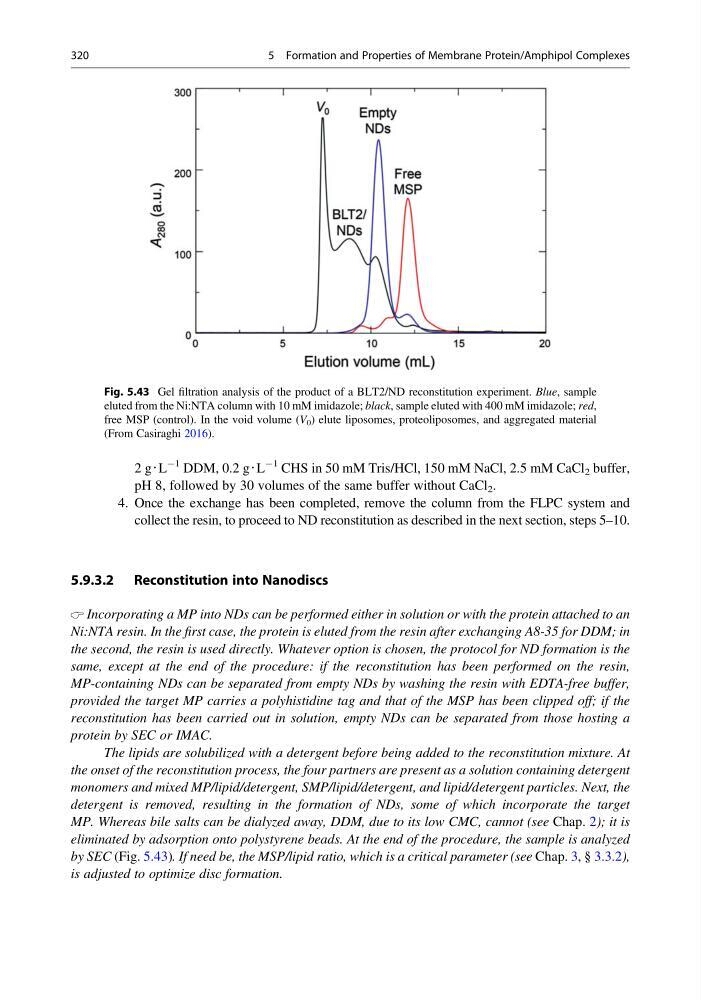
eliminated by adsorption onto polystyrene beads. At the end of the procedure, the sample is analyzed
by SEC (Fig. 5.43). If need be, the MSP/lipid ratio, which is a critical parameter (see Chap. 3, § 3.3.2),
is adjusted to optimize disc formation.
Fig. 5.43 Gel filtration analysis of the product of a BLT2/ND reconstitution experiment. Blue, sample
eluted from the Ni:NTA column with 10 mM imidazole; black, sample eluted with 400 mM imidazole; red,
free MSP (control). In the void volume (V 0 ) elute liposomes, proteoliposomes, and aggregated material
(From Casiraghi 2016).
320
5 Formation and Properties of Membrane Protein/Amphipol Complexes
À1 DDM, 0.2 g‧L
À1 CHS in 50 mM Tris/HCl, 150 mM NaCl, 2.5 mM CaCl 2 buffer,
pH 8, followed by 30 volumes of the same buffer without CaCl 2 .
4. Once the exchange has been completed, remove the column from the FLPC system and
collect the resin, to proceed to ND reconstitution as described in the next section, steps 5–10.
5.9.3.2 Reconstitution into Nanodiscs
Incorporating a MP into NDs can be performed either in solution or with the protein attached to an
Ni:NTA resin. In the first case, the protein is eluted from the resin after exchanging A8-35 for DDM; in
the second, the resin is used directly. Whatever option is chosen, the protocol for ND formation is the
same, except at the end of the procedure: if the reconstitution has been performed on the resin,
MP-containing NDs can be separated from empty NDs by washing the resin with EDTA-free buffer,
provided the target MP carries a polyhistidine tag and that of the MSP has been clipped off; if the
reconstitution has been carried out in solution, empty NDs can be separated from those hosting a
protein by SEC or IMAC.
The lipids are solubilized with a detergent before being added to the reconstitution mixture. At
the onset of the reconstitution process, the four partners are present as a solution containing detergent
monomers and mixed MP/lipid/detergent, SMP/lipid/detergent, and lipid/detergent particles. Next, the
detergent is removed, resulting in the formation of NDs, some of which incorporate the target
MP. Whereas bile salts can be dialyzed away, DDM, due to its low CMC, cannot (see Chap. 2); it is
eliminated by adsorption onto polystyrene beads. At the end of the procedure, the sample is analyzed
by SEC (Fig. 5.43). If need be, the MSP/lipid ratio, which is a critical parameter (see Chap. 3, § 3.3.2),
is adjusted to optimize disc formation.
Fig. 5.43 Gel filtration analysis of the product of a BLT2/ND reconstitution experiment. Blue, sample
eluted from the Ni:NTA column with 10 mM imidazole; black, sample eluted with 400 mM imidazole; red,
free MSP (control). In the void volume (V 0 ) elute liposomes, proteoliposomes, and aggregated material
(From Casiraghi 2016).
320
5 Formation and Properties of Membrane Protein/Amphipol Complexes