

unknown, its concentration can be assessed by colorimetric measurements such as bicinchoninic acid
(BCA) assay. Alternatively, amino acid analysis after HCl hydrolysis can also be used.
5.9.1.3 Determination of the Optimal MP/APol Mass Ratio
The sole optimization required is that of the mass ratio of APols needed to keep the MP soluble and
well dispersed in aqueous solution after detergent removal. For that, the protein and detergent
concentrations are kept unchanged, while increasing concentrations of APols are tested.
• Determine the range of MP/APol mass ratios to be tested: when starting on a novel MP, the
mass of APol each molecule will likely bind can be roughly estimated on the basis of the size
of the TM region (the MW of the protein being irrelevant). No precise relationship has been
worked out yet between the extent of the TM surface of the protein and the mass of APol that
will bind to it, but an estimate can be derived from the data in Table 5.5 and Fig. 5.9. A range
of 1Â to 10Â this estimate is a good starting point. If too little APol is used, aggregates will
form, which can be detected, crudely, by precipitation upon ultracentrifugation, and, in a
second stage and more precisely, by SEC. An excess of APol is useless and can be detrimental
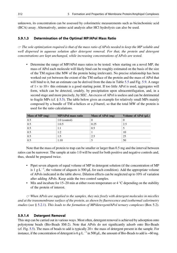
to fragile MPs (cf. § 5.5). The table below gives an example for relatively small MPs mainly
composed by a bundle of TM α-helices or a β-barrel, so that the total MW of the protein is
used for the ratio calculations.
Mass of MP (mg)
MP/APol mass ratio
Mass of APol (mg)
Volume of APol (μL)
0.5
1:0 (control)
0
0
0.5
1:0.5
0.25
2.5
0.5
1:1
0.5
5
0.5
1:2
1
10
0.5
1:5
2.5
25
0.5
1:10
5
50
Note that the mass of protein to trap can be smaller or larger than 0.5 mg and the interval between
ratios can be narrower. The sample at ratio 1:0 will be used for both positive and negative controls and,
thus, should be prepared twice.
• Pipet seven aliquots of equal volume of MP in detergent solution (if the concentration of MP
is 1 gÁL
À1 , the volume of aliquots is 500 μL for each condition). Add the appropriate volume
of APols indicated in the table above. Dilution effects can be neglected up to 10% of variation
after adding APols. Keep aside the two control samples.
• Mix and incubate for 15–20 min at either room temperature or 4
C depending on the stability
of the protein of interest.
When APols are supplied to the samples, they mix freely with detergent molecules in micelles
and at the transmembrane surface of the protein, as shown by fluorescence and isothermal calorimetry
studies (see § 5.2.1). This leads to the formation of MP/detergent/APol ternary complexes (Box 5.2).
5.9.1.4 Detergent Removal
This step can be carried out in various ways. Most often, detergent removal is achieved by adsorption onto
polystyrene beads (Bio-Beads SM-2). Note that APols do not significantly adsorb onto Bio-Beads
(cf. Fig. 5.5). The mass of beads to add is typically 20Â the mass of detergent present in the sample. For
instance, if the concentration of detergent is 6 gÁL
À1 in 500 μL, the amount of Bio-Beads to add is ~60 mg.
312
5 Formation and Properties of Membrane Protein/Amphipol Complexes
(BCA) assay. Alternatively, amino acid analysis after HCl hydrolysis can also be used.
5.9.1.3 Determination of the Optimal MP/APol Mass Ratio
The sole optimization required is that of the mass ratio of APols needed to keep the MP soluble and
well dispersed in aqueous solution after detergent removal. For that, the protein and detergent
concentrations are kept unchanged, while increasing concentrations of APols are tested.
• Determine the range of MP/APol mass ratios to be tested: when starting on a novel MP, the
mass of APol each molecule will likely bind can be roughly estimated on the basis of the size
of the TM region (the MW of the protein being irrelevant). No precise relationship has been
worked out yet between the extent of the TM surface of the protein and the mass of APol that
will bind to it, but an estimate can be derived from the data in Table 5.5 and Fig. 5.9. A range
of 1Â to 10Â this estimate is a good starting point. If too little APol is used, aggregates will
form, which can be detected, crudely, by precipitation upon ultracentrifugation, and, in a
second stage and more precisely, by SEC. An excess of APol is useless and can be detrimental
to fragile MPs (cf. § 5.5). The table below gives an example for relatively small MPs mainly
composed by a bundle of TM α-helices or a β-barrel, so that the total MW of the protein is
used for the ratio calculations.
Mass of MP (mg)
MP/APol mass ratio
Mass of APol (mg)
Volume of APol (μL)
0.5
1:0 (control)
0
0
0.5
1:0.5
0.25
2.5
0.5
1:1
0.5
5
0.5
1:2
1
10
0.5
1:5
2.5
25
0.5
1:10
5
50
Note that the mass of protein to trap can be smaller or larger than 0.5 mg and the interval between
ratios can be narrower. The sample at ratio 1:0 will be used for both positive and negative controls and,
thus, should be prepared twice.
• Pipet seven aliquots of equal volume of MP in detergent solution (if the concentration of MP
is 1 gÁL
À1 , the volume of aliquots is 500 μL for each condition). Add the appropriate volume
of APols indicated in the table above. Dilution effects can be neglected up to 10% of variation
after adding APols. Keep aside the two control samples.
• Mix and incubate for 15–20 min at either room temperature or 4
C depending on the stability
of the protein of interest.
When APols are supplied to the samples, they mix freely with detergent molecules in micelles
and at the transmembrane surface of the protein, as shown by fluorescence and isothermal calorimetry
studies (see § 5.2.1). This leads to the formation of MP/detergent/APol ternary complexes (Box 5.2).
5.9.1.4 Detergent Removal
This step can be carried out in various ways. Most often, detergent removal is achieved by adsorption onto
polystyrene beads (Bio-Beads SM-2). Note that APols do not significantly adsorb onto Bio-Beads
(cf. Fig. 5.5). The mass of beads to add is typically 20Â the mass of detergent present in the sample. For
instance, if the concentration of detergent is 6 gÁL
À1 in 500 μL, the amount of Bio-Beads to add is ~60 mg.
312
5 Formation and Properties of Membrane Protein/Amphipol Complexes