

chains or a mixture of apoA-I and apoA-II ones) are combined with a bilayer-forming mixture of lipids,
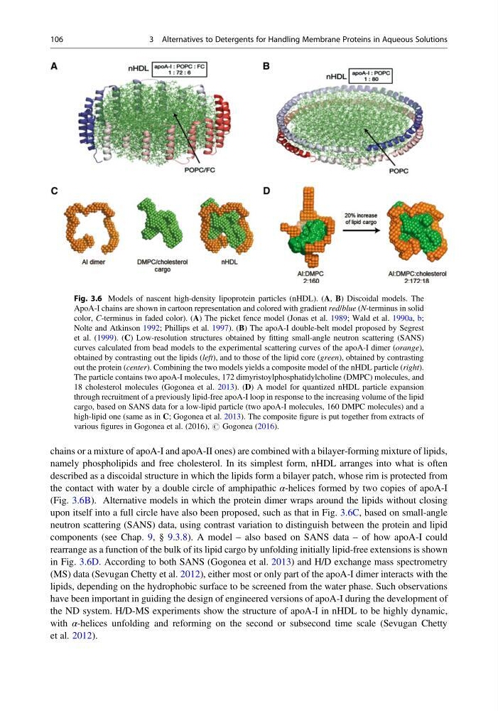
namely phospholipids and free cholesterol. In its simplest form, nHDL arranges into what is often
described as a discoidal structure in which the lipids form a bilayer patch, whose rim is protected from
the contact with water by a double circle of amphipathic α-helices formed by two copies of apoA-I
(Fig. 3.6B). Alternative models in which the protein dimer wraps around the lipids without closing
upon itself into a full circle have also been proposed, such as that in Fig. 3.6C, based on small-angle
neutron scattering (SANS) data, using contrast variation to distinguish between the protein and lipid
components (see Chap. 9, § 9.3.8). A model – also based on SANS data – of how apoA-I could
rearrange as a function of the bulk of its lipid cargo by unfolding initially lipid-free extensions is shown
in Fig. 3.6D. According to both SANS (Gogonea et al. 2013) and H/D exchange mass spectrometry
(MS) data (Sevugan Chetty et al. 2012), either most or only part of the apoA-I dimer interacts with the
lipids, depending on the hydrophobic surface to be screened from the water phase. Such observations
have been important in guiding the design of engineered versions of apoA-I during the development of
the ND system. H/D-MS experiments show the structure of apoA-I in nHDL to be highly dynamic,
with α-helices unfolding and reforming on the second or subsecond time scale (Sevugan Chetty
et al. 2012).
Fig. 3.6 Models of nascent high-density lipoprotein particles (nHDL). (A, B) Discoidal models. The
ApoA-I chains are shown in cartoon representation and colored with gradient red/blue (N-terminus in solid
color, C-terminus in faded color). (A) The picket fence model (Jonas et al. 1989; Wald et al. 1990a, b;
Nolte and Atkinson 1992; Phillips et al. 1997). (B) The apoA-I double-belt model proposed by Segrest
et al. (1999). (C) Low-resolution structures obtained by fitting small-angle neutron scattering (SANS)
curves calculated from bead models to the experimental scattering curves of the apoA-I dimer (orange),
obtained by contrasting out the lipids (left), and to those of the lipid core (green), obtained by contrasting
out the protein (center). Combining the two models yields a composite model of the nHDL particle (right).
The particle contains two apoA-I molecules, 172 dimyristoylphosphatidylcholine (DMPC) molecules, and
18 cholesterol molecules (Gogonea et al. 2013). (D) A model for quantized nHDL particle expansion
through recruitment of a previously lipid-free apoA-I loop in response to the increasing volume of the lipid
cargo, based on SANS data for a low-lipid particle (two apoA-I molecules, 160 DMPC molecules) and a
high-lipid one (same as in C; Gogonea et al. 2013). The composite figure is put together from extracts of
various figures in Gogonea et al. (2016), # Gogonea (2016).
106
3 Alternatives to Detergents for Handling Membrane Proteins in Aqueous Solutions
namely phospholipids and free cholesterol. In its simplest form, nHDL arranges into what is often
described as a discoidal structure in which the lipids form a bilayer patch, whose rim is protected from
the contact with water by a double circle of amphipathic α-helices formed by two copies of apoA-I
(Fig. 3.6B). Alternative models in which the protein dimer wraps around the lipids without closing
upon itself into a full circle have also been proposed, such as that in Fig. 3.6C, based on small-angle
neutron scattering (SANS) data, using contrast variation to distinguish between the protein and lipid
components (see Chap. 9, § 9.3.8). A model – also based on SANS data – of how apoA-I could
rearrange as a function of the bulk of its lipid cargo by unfolding initially lipid-free extensions is shown
in Fig. 3.6D. According to both SANS (Gogonea et al. 2013) and H/D exchange mass spectrometry
(MS) data (Sevugan Chetty et al. 2012), either most or only part of the apoA-I dimer interacts with the
lipids, depending on the hydrophobic surface to be screened from the water phase. Such observations
have been important in guiding the design of engineered versions of apoA-I during the development of
the ND system. H/D-MS experiments show the structure of apoA-I in nHDL to be highly dynamic,
with α-helices unfolding and reforming on the second or subsecond time scale (Sevugan Chetty
et al. 2012).
Fig. 3.6 Models of nascent high-density lipoprotein particles (nHDL). (A, B) Discoidal models. The
ApoA-I chains are shown in cartoon representation and colored with gradient red/blue (N-terminus in solid
color, C-terminus in faded color). (A) The picket fence model (Jonas et al. 1989; Wald et al. 1990a, b;
Nolte and Atkinson 1992; Phillips et al. 1997). (B) The apoA-I double-belt model proposed by Segrest
et al. (1999). (C) Low-resolution structures obtained by fitting small-angle neutron scattering (SANS)
curves calculated from bead models to the experimental scattering curves of the apoA-I dimer (orange),
obtained by contrasting out the lipids (left), and to those of the lipid core (green), obtained by contrasting
out the protein (center). Combining the two models yields a composite model of the nHDL particle (right).
The particle contains two apoA-I molecules, 172 dimyristoylphosphatidylcholine (DMPC) molecules, and
18 cholesterol molecules (Gogonea et al. 2013). (D) A model for quantized nHDL particle expansion
through recruitment of a previously lipid-free apoA-I loop in response to the increasing volume of the lipid
cargo, based on SANS data for a low-lipid particle (two apoA-I molecules, 160 DMPC molecules) and a
high-lipid one (same as in C; Gogonea et al. 2013). The composite figure is put together from extracts of
various figures in Gogonea et al. (2016), # Gogonea (2016).
106
3 Alternatives to Detergents for Handling Membrane Proteins in Aqueous Solutions