

Faham and James U. Bowie established, using as a model MP the usual victim of such forays into
unknown territories, bacteriorhodopsin (BR), that bicelles provide a favorable starting point for MP
crystallization (Faham and Bowie 2002; Faham et al. 2005). The approach was then extended to a
GPCR, the β 2 adrenergic receptor (Rasmussen et al. 2007), to the voltage-dependent anion channel 1
(Ujwal et al. 2008), to xanthorhodopsin (Luecke et al. 2008), to the rhomboid protease (Vinothkumar
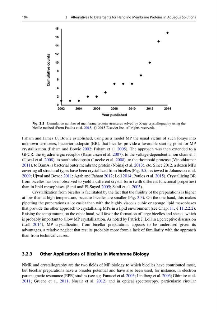
2011), to BamA, a bacterial outer membrane protein (Noinaj et al. 2013), etc. Since 2012, a dozen MPs
covering all structural types have been crystallized from bicelles (Fig. 3.5; reviewed in Johansson et al.
2009; Ujwal and Bowie 2011; Agah and Faham 2012; Loll 2014; Poulos et al. 2015). Crystallizing BR
from bicelles has been observed to yield a different crystal form (with different functional properties)
than in lipid mesophases (Sanii and El-Sayed 2005; Sanii et al. 2005).
Crystallization from bicelles is facilitated by the fact that the fluidity of the preparations is higher
at low than at high temperature, because bicelles are smaller (Fig. 3.3). On the one hand, this makes
pipetting the preparations a lot easier than with the highly viscous cubic or sponge lipid mesophases
that provide the other approach to crystallizing MPs in a lipid environment (see Chap. 11, § 11.2.2.2).
Raising the temperature, on the other hand, will favor the formation of large bicelles and sheets, which
is probably important to allow MP crystallization. As noted by Patrick J. Loll in a perceptive discussion
(Loll 2014), MP crystallization from bicellar preparations appears to be underused given its
advantages, a relative neglect that results probably more from a lack of familiarity with the approach
than from technical causes.
3.2.3
Other Applications of Bicelles in Membrane Biology
NMR and crystallography are the two fields of MP biology to which bicelles have contributed most,
but bicellar preparations have a broader potential and have also been used, for instance, in electron
paramagnetic resonance (EPR) studies (see e.g. Fanucci et al. 2003; Lindberg et al. 2003; Ghimire et al.
2011; Gruene et al. 2011; Nusair et al. 2012) and in optical spectroscopy, particularly circular
Fig. 3.5 Cumulative number of membrane protein structures solved by X-ray crystallography using the
bicelle method (From Poulos et al. 2015, # 2015 Elsevier Inc. All rights reserved).
104
3 Alternatives to Detergents for Handling Membrane Proteins in Aqueous Solutions
unknown territories, bacteriorhodopsin (BR), that bicelles provide a favorable starting point for MP
crystallization (Faham and Bowie 2002; Faham et al. 2005). The approach was then extended to a
GPCR, the β 2 adrenergic receptor (Rasmussen et al. 2007), to the voltage-dependent anion channel 1
(Ujwal et al. 2008), to xanthorhodopsin (Luecke et al. 2008), to the rhomboid protease (Vinothkumar
2011), to BamA, a bacterial outer membrane protein (Noinaj et al. 2013), etc. Since 2012, a dozen MPs
covering all structural types have been crystallized from bicelles (Fig. 3.5; reviewed in Johansson et al.
2009; Ujwal and Bowie 2011; Agah and Faham 2012; Loll 2014; Poulos et al. 2015). Crystallizing BR
from bicelles has been observed to yield a different crystal form (with different functional properties)
than in lipid mesophases (Sanii and El-Sayed 2005; Sanii et al. 2005).
Crystallization from bicelles is facilitated by the fact that the fluidity of the preparations is higher
at low than at high temperature, because bicelles are smaller (Fig. 3.3). On the one hand, this makes
pipetting the preparations a lot easier than with the highly viscous cubic or sponge lipid mesophases
that provide the other approach to crystallizing MPs in a lipid environment (see Chap. 11, § 11.2.2.2).
Raising the temperature, on the other hand, will favor the formation of large bicelles and sheets, which
is probably important to allow MP crystallization. As noted by Patrick J. Loll in a perceptive discussion
(Loll 2014), MP crystallization from bicellar preparations appears to be underused given its
advantages, a relative neglect that results probably more from a lack of familiarity with the approach
than from technical causes.
3.2.3
Other Applications of Bicelles in Membrane Biology
NMR and crystallography are the two fields of MP biology to which bicelles have contributed most,
but bicellar preparations have a broader potential and have also been used, for instance, in electron
paramagnetic resonance (EPR) studies (see e.g. Fanucci et al. 2003; Lindberg et al. 2003; Ghimire et al.
2011; Gruene et al. 2011; Nusair et al. 2012) and in optical spectroscopy, particularly circular
Fig. 3.5 Cumulative number of membrane protein structures solved by X-ray crystallography using the
bicelle method (From Poulos et al. 2015, # 2015 Elsevier Inc. All rights reserved).
104
3 Alternatives to Detergents for Handling Membrane Proteins in Aqueous Solutions