

g e , ¼2.002319304386. And Magnetic field for resonance is 3400 Gauss (0.34 Tesla)
for applied X-band microwave (9.5 GHz). There are two methods for observing ESR
signal at resonance condition. One is changing wavelength of microwave at constant
magnetic field and another is sweeping magnetic field at constant microwave. Most
of ESR machines adopt sweeping magnetic field method.
Stabilized nitroxide radicals are used as spin probes in spin labeling, and electron
spin of radical is spitted by hyperfine interaction (S・T・I) with quantized nuclear
spin of nitrogen atom (I¼À1, 0 and +1) (Fig. 3.7). The hyperfine interaction results
in three absorption peaks on ESR spectrum, and position of resonance absorption
and distance among three absorption lines on ESR spectrum are determined by
g-factor and T-tensor, respectively. And there is anisotropy caused by direction of
molecular axis because of localization of radical electron on πorbital of nitrogen
atom. When z-axis and x-axis are applied to direction of πorbital and direction of
N-O bond in molecular axis respectively. Anisotropic ESR spectra are obtained at
9.2 GHz microwave for each direction of applied magnetic field (Fig. 3.8).
3.6.1.1 Order Parameter, S
In spin probes of stearic acid and phosphatidylcholine, z-axis of the nitroxide
radicals coincide major axis of the molecules (Fig. 3.9). Molecular motion of these
molecules in lipid bilayers of biomembranes appears on ESR spectrum affected by
anisotropy of g-factor and T-tensor. When Z-axis of laboratory coordinate (X,Y,Z) is
applied to z-axis of molecular coordinate (x, y, z), each direction cosine of Z-axis
becomes cosα, cosβ and cosγ. In this case, ESR spectrum is obtained as time
averaged values of g-tensor and T-tensor in transformation from (x, y, z) coordinate
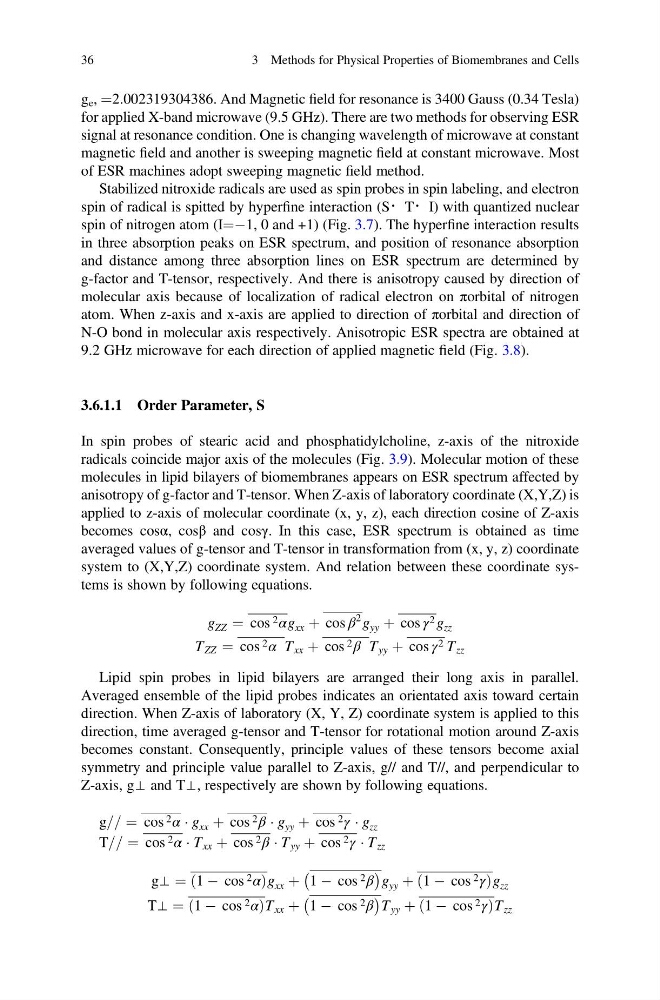
system to (X,Y,Z) coordinate system. And relation between these coordinate systems is shown by following equations.
g ZZ ¼ cos 2 αg xx þ cos β
2 g yy þ cos γ 2 g zz
T ZZ ¼ cos 2 α T xx þ cos 2 β T yy þ cos γ 2 T zz
Lipid spin probes in lipid bilayers are arranged their long axis in parallel.
Averaged ensemble of the lipid probes indicates an orientated axis toward certain
direction. When Z-axis of laboratory (X, Y, Z) coordinate system is applied to this
direction, time averaged g-tensor and T-tensor for rotational motion around Z-axis
becomes constant. Consequently, principle values of these tensors become axial
symmetry and principle value parallel to Z-axis, g// and T//, and perpendicular to
Z-axis, g⊥ and T⊥, respectively are shown by following equations.
g== ¼ cos 2 α Á g xx þ cos 2 β Á g yy þ cos 2 γ Á g zz
T== ¼ cos 2 α Á T xx þ cos 2 β Á T yy þ cos 2 γ Á T zz
g⊥ ¼ 1 À cos 2 α
ð
Þ g xx þ
À
1 À cos 2 β
Á
g yy þ 1 À cos 2 γ
ð
Þ g zz
T⊥ ¼ 1 À cos 2 α
ð
Þ T xx þ
À
1 À cos 2 β
Á
T yy þ 1 À cos 2 γ
ð
Þ T zz
36
3 Methods for Physical Properties of Biomembranes and Cells
for applied X-band microwave (9.5 GHz). There are two methods for observing ESR
signal at resonance condition. One is changing wavelength of microwave at constant
magnetic field and another is sweeping magnetic field at constant microwave. Most
of ESR machines adopt sweeping magnetic field method.
Stabilized nitroxide radicals are used as spin probes in spin labeling, and electron
spin of radical is spitted by hyperfine interaction (S・T・I) with quantized nuclear
spin of nitrogen atom (I¼À1, 0 and +1) (Fig. 3.7). The hyperfine interaction results
in three absorption peaks on ESR spectrum, and position of resonance absorption
and distance among three absorption lines on ESR spectrum are determined by
g-factor and T-tensor, respectively. And there is anisotropy caused by direction of
molecular axis because of localization of radical electron on πorbital of nitrogen
atom. When z-axis and x-axis are applied to direction of πorbital and direction of
N-O bond in molecular axis respectively. Anisotropic ESR spectra are obtained at
9.2 GHz microwave for each direction of applied magnetic field (Fig. 3.8).
3.6.1.1 Order Parameter, S
In spin probes of stearic acid and phosphatidylcholine, z-axis of the nitroxide
radicals coincide major axis of the molecules (Fig. 3.9). Molecular motion of these
molecules in lipid bilayers of biomembranes appears on ESR spectrum affected by
anisotropy of g-factor and T-tensor. When Z-axis of laboratory coordinate (X,Y,Z) is
applied to z-axis of molecular coordinate (x, y, z), each direction cosine of Z-axis
becomes cosα, cosβ and cosγ. In this case, ESR spectrum is obtained as time
averaged values of g-tensor and T-tensor in transformation from (x, y, z) coordinate
system to (X,Y,Z) coordinate system. And relation between these coordinate systems is shown by following equations.
g ZZ ¼ cos 2 αg xx þ cos β
2 g yy þ cos γ 2 g zz
T ZZ ¼ cos 2 α T xx þ cos 2 β T yy þ cos γ 2 T zz
Lipid spin probes in lipid bilayers are arranged their long axis in parallel.
Averaged ensemble of the lipid probes indicates an orientated axis toward certain
direction. When Z-axis of laboratory (X, Y, Z) coordinate system is applied to this
direction, time averaged g-tensor and T-tensor for rotational motion around Z-axis
becomes constant. Consequently, principle values of these tensors become axial
symmetry and principle value parallel to Z-axis, g// and T//, and perpendicular to
Z-axis, g⊥ and T⊥, respectively are shown by following equations.
g== ¼ cos 2 α Á g xx þ cos 2 β Á g yy þ cos 2 γ Á g zz
T== ¼ cos 2 α Á T xx þ cos 2 β Á T yy þ cos 2 γ Á T zz
g⊥ ¼ 1 À cos 2 α
ð
Þ g xx þ
À
1 À cos 2 β
Á
g yy þ 1 À cos 2 γ
ð
Þ g zz
T⊥ ¼ 1 À cos 2 α
ð
Þ T xx þ
À
1 À cos 2 β
Á
T yy þ 1 À cos 2 γ
ð
Þ T zz
36
3 Methods for Physical Properties of Biomembranes and Cells