

Position of electron is maximum position of electron density in deed. Electron
density at position coordinate (x,y,z) in the unit lattice is shown by
ρ xyz
ð Þ ¼
X h
Àh
X k
Àk
X l
Àl
F hkl
ð Þe
À2πi
À
hx j þky j þlz j . Although structure factor is Fourier
transformation, this equation is inverse Fourier transformation. Only scattering
intensity |F(hkl)| is obtained by X-ray diffraction data, and relationship between
the scattering intensity and X-ray diffraction is shown by F(hkl) ¼ |F(hkl)e|
À2πiφ ,
where φ is phase. To obtain electron density, phase should be known. It is necessary
to infer phase in X-ray crystallography. There are direct method and heavy atom
replacement method to infer phase of X-ray diffraction.
3.3 Electron Microscopy
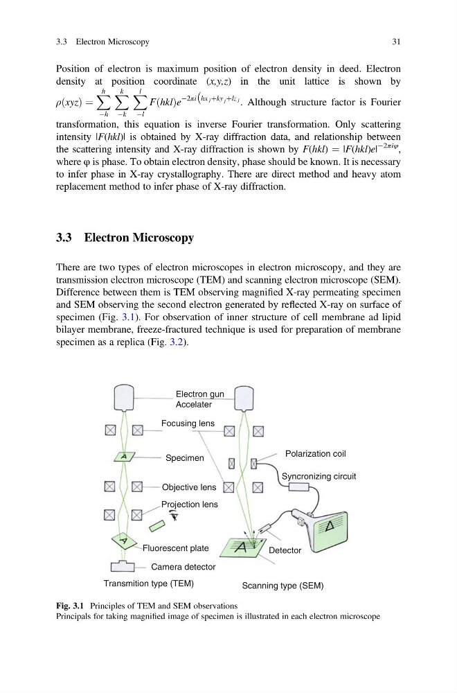
There are two types of electron microscopes in electron microscopy, and they are
transmission electron microscope (TEM) and scanning electron microscope (SEM).
Difference between them is TEM observing magnified X-ray permeating specimen
and SEM observing the second electron generated by reflected X-ray on surface of
specimen (Fig. 3.1). For observation of inner structure of cell membrane ad lipid
bilayer membrane, freeze-fractured technique is used for preparation of membrane
specimen as a replica (Fig. 3.2).
Electron gun
Accelater
Focusing lens
Specimen
Polarization coil
Syncronizing circuit
Detector
Fluorescent plate
Camera detector
Objective lens
Projection lens
Transmition type (TEM)
Scanning type (SEM)
Fig. 3.1 Principles of TEM and SEM observations
Principals for taking magnified image of specimen is illustrated in each electron microscope
3.3 Electron Microscopy
31
density at position coordinate (x,y,z) in the unit lattice is shown by
ρ xyz
ð Þ ¼
X h
Àh
X k
Àk
X l
Àl
F hkl
ð Þe
À2πi
À
hx j þky j þlz j . Although structure factor is Fourier
transformation, this equation is inverse Fourier transformation. Only scattering
intensity |F(hkl)| is obtained by X-ray diffraction data, and relationship between
the scattering intensity and X-ray diffraction is shown by F(hkl) ¼ |F(hkl)e|
À2πiφ ,
where φ is phase. To obtain electron density, phase should be known. It is necessary
to infer phase in X-ray crystallography. There are direct method and heavy atom
replacement method to infer phase of X-ray diffraction.
3.3 Electron Microscopy
There are two types of electron microscopes in electron microscopy, and they are
transmission electron microscope (TEM) and scanning electron microscope (SEM).
Difference between them is TEM observing magnified X-ray permeating specimen
and SEM observing the second electron generated by reflected X-ray on surface of
specimen (Fig. 3.1). For observation of inner structure of cell membrane ad lipid
bilayer membrane, freeze-fractured technique is used for preparation of membrane
specimen as a replica (Fig. 3.2).
Electron gun
Accelater
Focusing lens
Specimen
Polarization coil
Syncronizing circuit
Detector
Fluorescent plate
Camera detector
Objective lens
Projection lens
Transmition type (TEM)
Scanning type (SEM)
Fig. 3.1 Principles of TEM and SEM observations
Principals for taking magnified image of specimen is illustrated in each electron microscope
3.3 Electron Microscopy
31