

to dissociate two disulfide bonds of RNase A in 8M urea solution. The denatured
RNase A was renatured by oxidative treatment of removing urea by dialysis at pH 8.
2.6 Cell as a Device of Information Processing
A cell receives external stimulus and coverts it to a signal that is available in a signal
transducing mechanism in the cell, and the cell shows a cellular function
corresponding to the stimulus. So, cell can be considered as an information
processing devise. There is a receptor protein on surface of a cell and it receives a
ligand (stimulus-molecule) specifically by complementary recognition of molecular
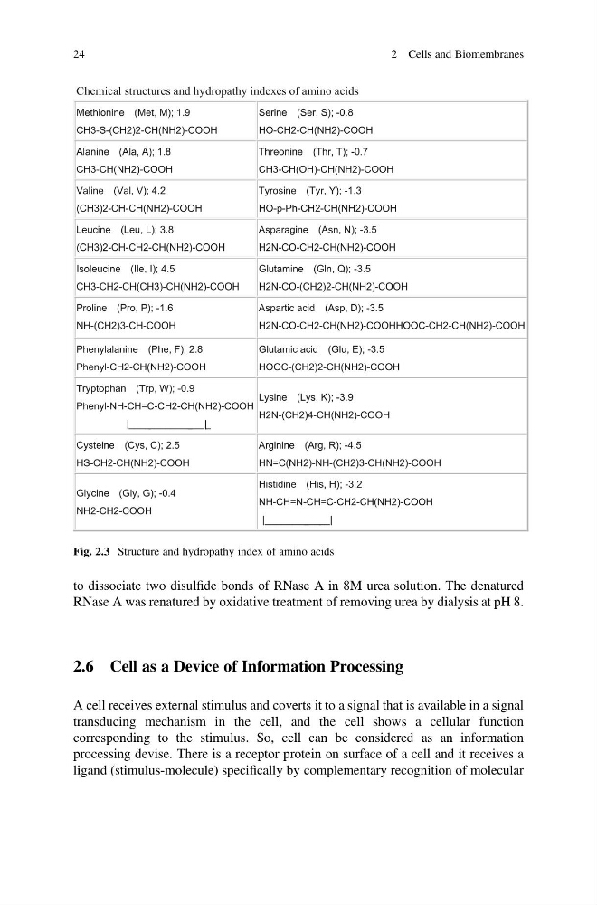
Chemical structures and hydropathy indexes of amino acids
Methionine (Met, M); 1.9
CH3-S-(CH2)2-CH(NH2)-COOH
Serine (Ser, S); -0.8
HO-CH2-CH(NH2)-COOH
Alanine (Ala, A); 1.8
CH3-CH(NH2)-COOH
Threonine (Thr, T); -0.7
CH3-CH(OH)-CH(NH2)-COOH
Valine (Val, V); 4.2
(CH3)2-CH-CH(NH2)-COOH
Tyrosine (Tyr, Y); -1.3
HO-p-Ph-CH2-CH(NH2)-COOH
Leucine (Leu, L); 3.8
(CH3)2-CH-CH2-CH(NH2)-COOH
Asparagine (Asn, N); -3.5
H2N-CO-CH2-CH(NH2)-COOH
Isoleucine (Ile, I); 4.5
CH3-CH2-CH(CH3)-CH(NH2)-COOH
Glutamine (Gln, Q); -3.5
H2N-CO-(CH2)2-CH(NH2)-COOH
Proline (Pro, P); -1.6
NH-(CH2)3-CH-COOH
Aspartic acid (Asp, D); -3.5
H2N-CO-CH2-CH(NH2)-COOHHOOC-CH2-CH(NH2)-COOH
Phenylalanine (Phe, F); 2.8
Phenyl-CH2-CH(NH2)-COOH
Glutamic acid (Glu, E); -3.5
HOOC-(CH2)2-CH(NH2)-COOH
Tryptophan (Trp, W); -0.9
Phenyl-NH-CH=C-CH2-CH(NH2)-COOH
|____________ ___| _
Lysine (Lys, K); -3.9
H2N-(CH2)4-CH(NH2)-COOH
Cysteine (Cys, C); 2.5
HS-CH2-CH(NH2)-COOH
Arginine (Arg, R); -4.5
HN=C(NH2)-NH-(CH2)3-CH(NH2)-COOH
Glycine (Gly, G); -0.4
NH2-CH2-COOH
Histidine (His, H); -3.2
NH-CH=N-CH=C-CH2-CH(NH2)-COOH
|________ _____|
Fig. 2.3 Structure and hydropathy index of amino acids
24
2 Cells and Biomembranes
RNase A was renatured by oxidative treatment of removing urea by dialysis at pH 8.
2.6 Cell as a Device of Information Processing
A cell receives external stimulus and coverts it to a signal that is available in a signal
transducing mechanism in the cell, and the cell shows a cellular function
corresponding to the stimulus. So, cell can be considered as an information
processing devise. There is a receptor protein on surface of a cell and it receives a
ligand (stimulus-molecule) specifically by complementary recognition of molecular
Chemical structures and hydropathy indexes of amino acids
Methionine (Met, M); 1.9
CH3-S-(CH2)2-CH(NH2)-COOH
Serine (Ser, S); -0.8
HO-CH2-CH(NH2)-COOH
Alanine (Ala, A); 1.8
CH3-CH(NH2)-COOH
Threonine (Thr, T); -0.7
CH3-CH(OH)-CH(NH2)-COOH
Valine (Val, V); 4.2
(CH3)2-CH-CH(NH2)-COOH
Tyrosine (Tyr, Y); -1.3
HO-p-Ph-CH2-CH(NH2)-COOH
Leucine (Leu, L); 3.8
(CH3)2-CH-CH2-CH(NH2)-COOH
Asparagine (Asn, N); -3.5
H2N-CO-CH2-CH(NH2)-COOH
Isoleucine (Ile, I); 4.5
CH3-CH2-CH(CH3)-CH(NH2)-COOH
Glutamine (Gln, Q); -3.5
H2N-CO-(CH2)2-CH(NH2)-COOH
Proline (Pro, P); -1.6
NH-(CH2)3-CH-COOH
Aspartic acid (Asp, D); -3.5
H2N-CO-CH2-CH(NH2)-COOHHOOC-CH2-CH(NH2)-COOH
Phenylalanine (Phe, F); 2.8
Phenyl-CH2-CH(NH2)-COOH
Glutamic acid (Glu, E); -3.5
HOOC-(CH2)2-CH(NH2)-COOH
Tryptophan (Trp, W); -0.9
Phenyl-NH-CH=C-CH2-CH(NH2)-COOH
|____________ ___| _
Lysine (Lys, K); -3.9
H2N-(CH2)4-CH(NH2)-COOH
Cysteine (Cys, C); 2.5
HS-CH2-CH(NH2)-COOH
Arginine (Arg, R); -4.5
HN=C(NH2)-NH-(CH2)3-CH(NH2)-COOH
Glycine (Gly, G); -0.4
NH2-CH2-COOH
Histidine (His, H); -3.2
NH-CH=N-CH=C-CH2-CH(NH2)-COOH
|________ _____|
Fig. 2.3 Structure and hydropathy index of amino acids
24
2 Cells and Biomembranes