

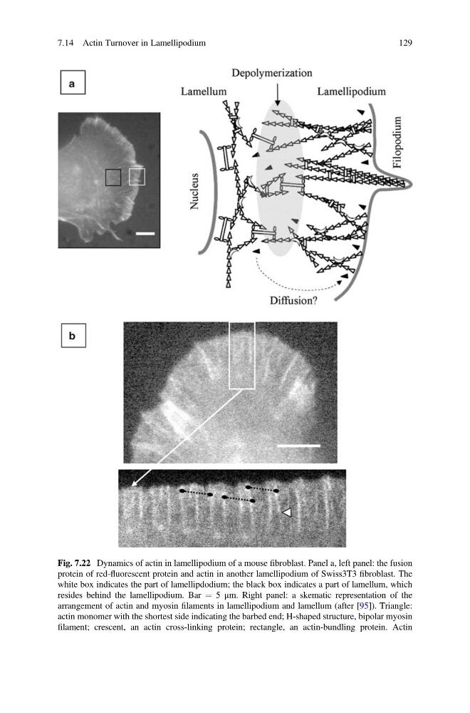
Fig. 7.22 Dynamics of actin in lamellipodium of a mouse fibroblast. Panel a, left panel: the fusion
protein of red-fluorescent protein and actin in another lamellipodium of Swiss3T3 fibroblast. The
white box indicates the part of lamellipdodium; the black box indicates a part of lamellum, which
resides behind the lamellipodium. Bar ¼ 5 μm. Right panel: a skematic representation of the
arrangement of actin and myosin filaments in lamellipodium and lamellum (after [95]). Triangle:
actin monomer with the shortest side indicating the barbed end; H-shaped structure, bipolar myosin
filament; crescent, an actin cross-linking protein; rectangle, an actin-bundling protein. Actin
7.14 Actin Turnover in Lamellipodium
129
protein of red-fluorescent protein and actin in another lamellipodium of Swiss3T3 fibroblast. The
white box indicates the part of lamellipdodium; the black box indicates a part of lamellum, which
resides behind the lamellipodium. Bar ¼ 5 μm. Right panel: a skematic representation of the
arrangement of actin and myosin filaments in lamellipodium and lamellum (after [95]). Triangle:
actin monomer with the shortest side indicating the barbed end; H-shaped structure, bipolar myosin
filament; crescent, an actin cross-linking protein; rectangle, an actin-bundling protein. Actin
7.14 Actin Turnover in Lamellipodium
129