

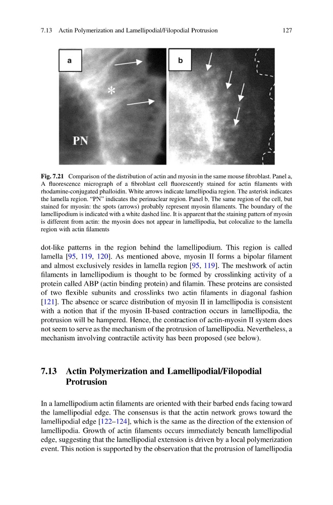
dot-like patterns in the region behind the lamellipodium. This region is called
lamella [95, 119, 120]. As mentioned above, myosin II forms a bipolar filament
and almost exclusively resides in lamella region [95, 119]. The meshwork of actin
filaments in lamellipodium is thought to be formed by crosslinking activity of a
protein called ABP (actin binding protein) and filamin. These proteins are consisted
of two flexible subunits and crosslinks two actin filaments in diagonal fashion
[121]. The absence or scarce distribution of myosin II in lamellipodia is consistent
with a notion that if the myosin II-based contraction occurs in lamellipodia, the
protrusion will be hampered. Hence, the contraction of actin-myosin II system does
not seem to serve as the mechanism of the protrusion of lamellipodia. Nevertheless, a
mechanism involving contractile activity has been proposed (see below).
7.13 Actin Polymerization and Lamellipodial/Filopodial
Protrusion
In a lamellipodium actin filaments are oriented with their barbed ends facing toward
the lamellipodial edge. The consensus is that the actin network grows toward the
lamellipodial edge [122–124], which is the same as the direction of the extension of
lamellipodia. Growth of actin filaments occurs immediately beneath lamellipodial
edge, suggesting that the lamellipodial extension is driven by a local polymerization
event. This notion is supported by the observation that the protrusion of lamellipodia
Fig. 7.21 Comparison of the distribution of actin and myosin in the same mouse fibroblast. Panel a,
A fluorescence micrograph of a fibroblast cell fluorescently stained for actin filaments with
rhodamine-conjugated phalloidin. White arrows indicate lamellipodia region. The asterisk indicates
the lamella region. “PN” indicates the perinuclear region. Panel b, The same region of the cell, but
stained for myosin: the spots (arrows) probably represent myosin filaments. The boundary of the
lamellipodium is indicated with a white dashed line. It is apparent that the staining pattern of myosin
is different from actin: the myosin does not appear in lamellipodia, but colocalize to the lamella
region with actin filaments
7.13 Actin Polymerization and Lamellipodial/Filopodial Protrusion
127
lamella [95, 119, 120]. As mentioned above, myosin II forms a bipolar filament
and almost exclusively resides in lamella region [95, 119]. The meshwork of actin
filaments in lamellipodium is thought to be formed by crosslinking activity of a
protein called ABP (actin binding protein) and filamin. These proteins are consisted
of two flexible subunits and crosslinks two actin filaments in diagonal fashion
[121]. The absence or scarce distribution of myosin II in lamellipodia is consistent
with a notion that if the myosin II-based contraction occurs in lamellipodia, the
protrusion will be hampered. Hence, the contraction of actin-myosin II system does
not seem to serve as the mechanism of the protrusion of lamellipodia. Nevertheless, a
mechanism involving contractile activity has been proposed (see below).
7.13 Actin Polymerization and Lamellipodial/Filopodial
Protrusion
In a lamellipodium actin filaments are oriented with their barbed ends facing toward
the lamellipodial edge. The consensus is that the actin network grows toward the
lamellipodial edge [122–124], which is the same as the direction of the extension of
lamellipodia. Growth of actin filaments occurs immediately beneath lamellipodial
edge, suggesting that the lamellipodial extension is driven by a local polymerization
event. This notion is supported by the observation that the protrusion of lamellipodia
Fig. 7.21 Comparison of the distribution of actin and myosin in the same mouse fibroblast. Panel a,
A fluorescence micrograph of a fibroblast cell fluorescently stained for actin filaments with
rhodamine-conjugated phalloidin. White arrows indicate lamellipodia region. The asterisk indicates
the lamella region. “PN” indicates the perinuclear region. Panel b, The same region of the cell, but
stained for myosin: the spots (arrows) probably represent myosin filaments. The boundary of the
lamellipodium is indicated with a white dashed line. It is apparent that the staining pattern of myosin
is different from actin: the myosin does not appear in lamellipodia, but colocalize to the lamella
region with actin filaments
7.13 Actin Polymerization and Lamellipodial/Filopodial Protrusion
127