

7.8 Extracelluar Matrix and Its Connection to Cytoskeleton
Cells produce and secrete proteins that form an extracellular network structure called
extracellular matrix (ECM; [80]). The ECM serves as the basis for the adhesion of
the cell to the surrounding cells or tissues, or structures such as the surface of a
culture dish. The binding of the cell to ECM is mediated by integrin, a family of
transmembrane protein. Integrin is a heterodimer of alpha- and beta-integrins. There
are many different alpha and beta integrins, and hence, there are a number of
combinations of alpha and beta-integrins. There are also many types of ECMs; for
example, fibronectin, collagen and laminin. The combination of alpha- and betaintegrin basically determines the partner ECM, for example alpha-5 beta-1 integrin
heterodimer binds to fibronectin [81]. Interestingly, specific combination of alphaand beta-integrins determines the partner cytoplasmic proteins that interact with the
integrin heterodimer [82]. Thus, the integrin heterodimer bidirectionally transmit the
information of the extracelluar binding event and the intracellular events
[83, 84]. The ECM-cytoskeleton connection plays an essential role when the cell
attaches its front part to substrate to pull the rear part by contraction.
7.9 Cell Membrane and Membrane Skeleton
As described in Chap. 4, the basis of the cell membrane is a lipid bilayer. The lipid
bilayer itself is a highly flexible entity: its bending rigidity is on the order of 10
-19 J,
allowing thermal fluctuations [85]. It should be noted that the tensile strength of the
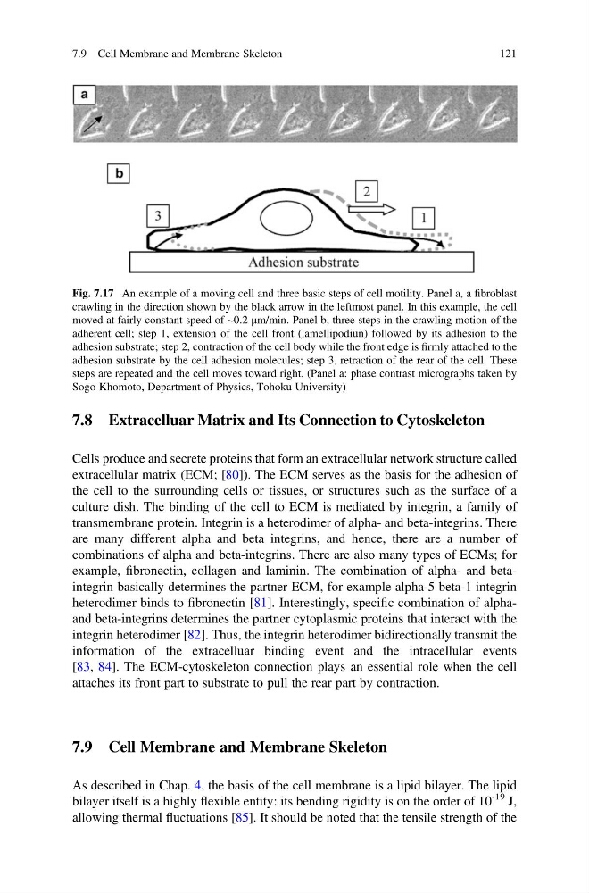
Fig. 7.17 An example of a moving cell and three basic steps of cell motility. Panel a, a fibroblast
crawling in the direction shown by the black arrow in the leftmost panel. In this example, the cell
moved at fairly constant speed of ~0.2 μm/min. Panel b, three steps in the crawling motion of the
adherent cell; step 1, extension of the cell front (lamellipodiun) followed by its adhesion to the
adhesion substrate; step 2, contraction of the cell body while the front edge is firmly attached to the
adhesion substrate by the cell adhesion molecules; step 3, retraction of the rear of the cell. These
steps are repeated and the cell moves toward right. (Panel a: phase contrast micrographs taken by
Sogo Khomoto, Department of Physics, Tohoku University)
7.9 Cell Membrane and Membrane Skeleton
121
Cells produce and secrete proteins that form an extracellular network structure called
extracellular matrix (ECM; [80]). The ECM serves as the basis for the adhesion of
the cell to the surrounding cells or tissues, or structures such as the surface of a
culture dish. The binding of the cell to ECM is mediated by integrin, a family of
transmembrane protein. Integrin is a heterodimer of alpha- and beta-integrins. There
are many different alpha and beta integrins, and hence, there are a number of
combinations of alpha and beta-integrins. There are also many types of ECMs; for
example, fibronectin, collagen and laminin. The combination of alpha- and betaintegrin basically determines the partner ECM, for example alpha-5 beta-1 integrin
heterodimer binds to fibronectin [81]. Interestingly, specific combination of alphaand beta-integrins determines the partner cytoplasmic proteins that interact with the
integrin heterodimer [82]. Thus, the integrin heterodimer bidirectionally transmit the
information of the extracelluar binding event and the intracellular events
[83, 84]. The ECM-cytoskeleton connection plays an essential role when the cell
attaches its front part to substrate to pull the rear part by contraction.
7.9 Cell Membrane and Membrane Skeleton
As described in Chap. 4, the basis of the cell membrane is a lipid bilayer. The lipid
bilayer itself is a highly flexible entity: its bending rigidity is on the order of 10
-19 J,
allowing thermal fluctuations [85]. It should be noted that the tensile strength of the
Fig. 7.17 An example of a moving cell and three basic steps of cell motility. Panel a, a fibroblast
crawling in the direction shown by the black arrow in the leftmost panel. In this example, the cell
moved at fairly constant speed of ~0.2 μm/min. Panel b, three steps in the crawling motion of the
adherent cell; step 1, extension of the cell front (lamellipodiun) followed by its adhesion to the
adhesion substrate; step 2, contraction of the cell body while the front edge is firmly attached to the
adhesion substrate by the cell adhesion molecules; step 3, retraction of the rear of the cell. These
steps are repeated and the cell moves toward right. (Panel a: phase contrast micrographs taken by
Sogo Khomoto, Department of Physics, Tohoku University)
7.9 Cell Membrane and Membrane Skeleton
121