

7.3.2 Actin Filament Structure
Actin monomers polymerize to form a long, flexible filament. As mentioned above,
we call the actin monomer incorporated in the filament actin “protomer”. The actin
filament looks like a right-handed, two-start helix according to the electron micrograph (Fig. 7.11a; [29]), but one can trace the individual monomers in the filament
along a left-handed, one-start helix [30]: the rise of the one-start helix is 2.75 nm and
the monomer makes ~166 degrees rotation around the filament axis: this nearly
180 degrees’ rotation of monomer makes the actin filament have the appearance of
the right-handed, two-start helix [28]: the pitch of this helix is 72 nm and the interprotomer distance along the filament axis is 5.5 nm. The two ends of filament can be
distinguished based on the difference in structural and kinetic properties concerning
the binding of the terminal protomers. The two, non-identical ends are named
“barbed” and “pointed” ends, according to the polarity as indicated by the bound
heavy meromyosin (see below).
The structure of actin filament at the atomic resolution was obtained from the
fitting of the filament model constructed from the crystal structure of monomer to an
X-ray diagram of actin gel oriented in a glass capillary [31]. The constructed model
exhibited that the subdomains had slightly change their positions upon the incorporation of the monomer into the filament. The subdomain II and IV of the last
protomer is exposed at the pointed end, while subdomain I and III are exposed at
the barbed end (Fig. 7.11b). The cleft between the large and the small domain is
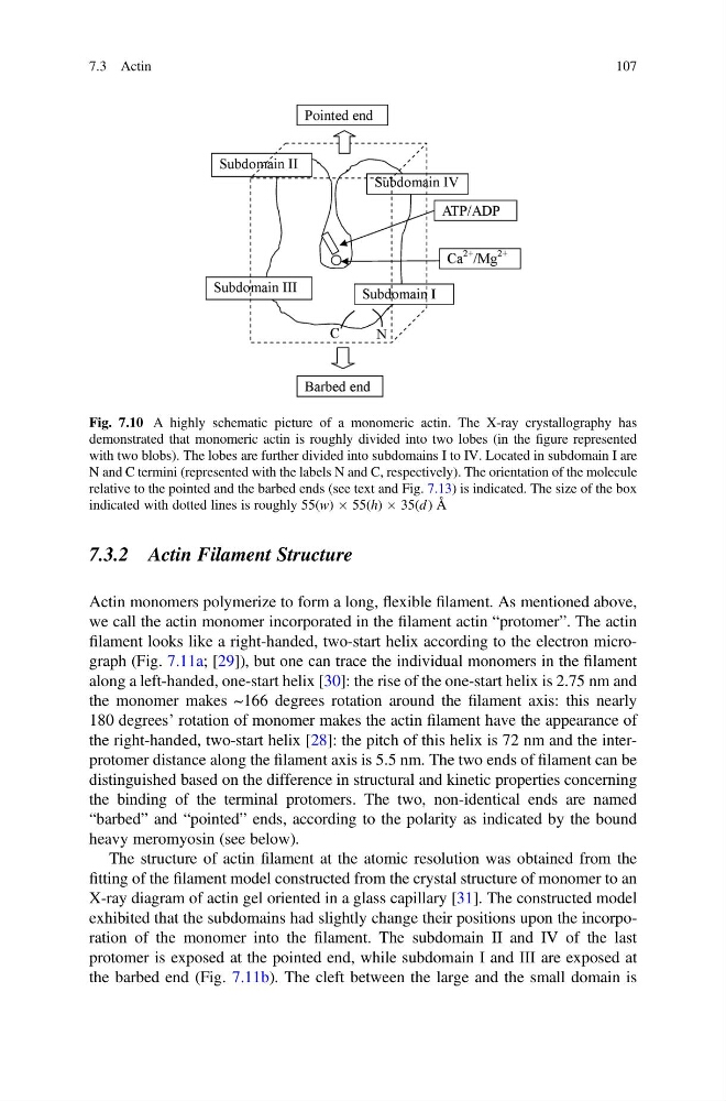
Fig. 7.10 A highly schematic picture of a monomeric actin. The X-ray crystallography has
demonstrated that monomeric actin is roughly divided into two lobes (in the figure represented
with two blobs). The lobes are further divided into subdomains I to IV. Located in subdomain I are
N and C termini (represented with the labels N and C, respectively). The orientation of the molecule
relative to the pointed and the barbed ends (see text and Fig. 7.13) is indicated. The size of the box
indicated with dotted lines is roughly 55(w) Â 55(h) Â 35(d ) Å
7.3 Actin
107
Actin monomers polymerize to form a long, flexible filament. As mentioned above,
we call the actin monomer incorporated in the filament actin “protomer”. The actin
filament looks like a right-handed, two-start helix according to the electron micrograph (Fig. 7.11a; [29]), but one can trace the individual monomers in the filament
along a left-handed, one-start helix [30]: the rise of the one-start helix is 2.75 nm and
the monomer makes ~166 degrees rotation around the filament axis: this nearly
180 degrees’ rotation of monomer makes the actin filament have the appearance of
the right-handed, two-start helix [28]: the pitch of this helix is 72 nm and the interprotomer distance along the filament axis is 5.5 nm. The two ends of filament can be
distinguished based on the difference in structural and kinetic properties concerning
the binding of the terminal protomers. The two, non-identical ends are named
“barbed” and “pointed” ends, according to the polarity as indicated by the bound
heavy meromyosin (see below).
The structure of actin filament at the atomic resolution was obtained from the
fitting of the filament model constructed from the crystal structure of monomer to an
X-ray diagram of actin gel oriented in a glass capillary [31]. The constructed model
exhibited that the subdomains had slightly change their positions upon the incorporation of the monomer into the filament. The subdomain II and IV of the last
protomer is exposed at the pointed end, while subdomain I and III are exposed at
the barbed end (Fig. 7.11b). The cleft between the large and the small domain is
Fig. 7.10 A highly schematic picture of a monomeric actin. The X-ray crystallography has
demonstrated that monomeric actin is roughly divided into two lobes (in the figure represented
with two blobs). The lobes are further divided into subdomains I to IV. Located in subdomain I are
N and C termini (represented with the labels N and C, respectively). The orientation of the molecule
relative to the pointed and the barbed ends (see text and Fig. 7.13) is indicated. The size of the box
indicated with dotted lines is roughly 55(w) Â 55(h) Â 35(d ) Å
7.3 Actin
107