

In this method, an aqueous solution of (NH 4 ) 2 HPO 4 was slowly added drop by drop
into aqueous Ca(NO 3 ) 2 solution under stirring at room temperature. The resultant
precipitate was put into an autoclave and then treated hydrothermally at 140
C for
5 h under a pressure of 0.3 MPa. It was then washed with deionized water by a
centrifugal method. Finally, the white slurry was dried to produce HAp [55].
Needle-like HAp nanocrystals having a similar morphology and crystal structure
to natural apatite (average size of 50 Â 25 nm and thickness of 2–3 nm) have been
synthesized by hydrothermal methods [54].
Slosarczyk et al. have used a wet method to obtain carbonated HAp powders [56,
57]. Calcium oxide (CaO), calcium nitrate, calcium tetrahydrate [Ca(NO 3 ) 2 ·4H 2 O]
or calcium acetate [Ca(CH 3 COO) 2 ·H 2 O] were used as the calcium source. As the
phosphorous source, phosphoric acid (H 3 PO 4 ) or di-ammonium phosphate
[(NH 4 ) 2 HPO 4 ] were used. The molar ratio of Ca:P was 1.67. Ammonium bicarbonate (NH 4 HCO 3 ) or sodium bicarbonate (NaHCO 3 ) were used as reactants to
introduce CO 3
2À groups. Biological apatites in natural bone, dentin, and enamel
contain 7.4, 5.6, and 3.5 wt% of carbonate, respectively. In this method, some of the
PO 4
3À and OH
À groups are replaced by CO 3
2À groups. This carbonated HAp
powder has proved to be a promising material for bioresorbable bone substitution,
among other applications [56].
Template technology has been used by Zhang et al. to synthesize HAp singlecrystal nanowires, and the product was confirmed by using different analytical
techniques, e.g., X-ray diffraction (XRD), transmission electron microscopy
(TEM), and X-ray photoelectron spectroscopy (XPS). The crystalline order of the
HAp precursors was maintained in the electrodeposited nanowires, which were
confirmed by the characterization. These HAp single-crystal nanowires displayed
structural similarity to the natural HAp found in bone [58].
The precipitation method was adopted by Siddharthan et al. to synthesize
calcium-deficient HAp (cdHAp) with a Ca:P ratio of 1.5 [53] using calcium nitrate
tetrahydrate and phosphoric acid under accelerated microwave irradiation. The
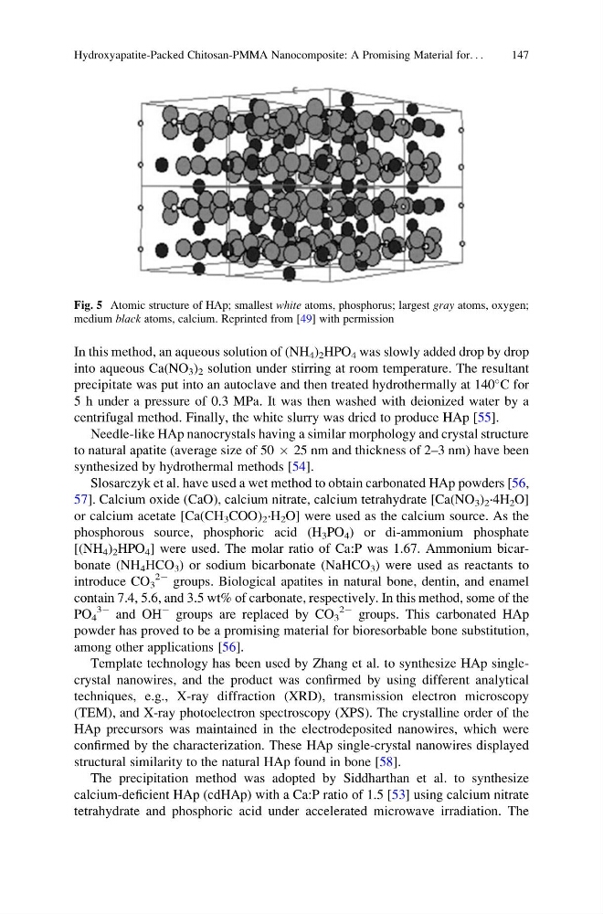
Fig. 5 Atomic structure of HAp; smallest white atoms, phosphorus; largest gray atoms, oxygen;
medium black atoms, calcium. Reprinted from [49] with permission
Hydroxyapatite-Packed Chitosan-PMMA Nanocomposite: A Promising Material for. . .
147
into aqueous Ca(NO 3 ) 2 solution under stirring at room temperature. The resultant
precipitate was put into an autoclave and then treated hydrothermally at 140
C for
5 h under a pressure of 0.3 MPa. It was then washed with deionized water by a
centrifugal method. Finally, the white slurry was dried to produce HAp [55].
Needle-like HAp nanocrystals having a similar morphology and crystal structure
to natural apatite (average size of 50 Â 25 nm and thickness of 2–3 nm) have been
synthesized by hydrothermal methods [54].
Slosarczyk et al. have used a wet method to obtain carbonated HAp powders [56,
57]. Calcium oxide (CaO), calcium nitrate, calcium tetrahydrate [Ca(NO 3 ) 2 ·4H 2 O]
or calcium acetate [Ca(CH 3 COO) 2 ·H 2 O] were used as the calcium source. As the
phosphorous source, phosphoric acid (H 3 PO 4 ) or di-ammonium phosphate
[(NH 4 ) 2 HPO 4 ] were used. The molar ratio of Ca:P was 1.67. Ammonium bicarbonate (NH 4 HCO 3 ) or sodium bicarbonate (NaHCO 3 ) were used as reactants to
introduce CO 3
2À groups. Biological apatites in natural bone, dentin, and enamel
contain 7.4, 5.6, and 3.5 wt% of carbonate, respectively. In this method, some of the
PO 4
3À and OH
À groups are replaced by CO 3
2À groups. This carbonated HAp
powder has proved to be a promising material for bioresorbable bone substitution,
among other applications [56].
Template technology has been used by Zhang et al. to synthesize HAp singlecrystal nanowires, and the product was confirmed by using different analytical
techniques, e.g., X-ray diffraction (XRD), transmission electron microscopy
(TEM), and X-ray photoelectron spectroscopy (XPS). The crystalline order of the
HAp precursors was maintained in the electrodeposited nanowires, which were
confirmed by the characterization. These HAp single-crystal nanowires displayed
structural similarity to the natural HAp found in bone [58].
The precipitation method was adopted by Siddharthan et al. to synthesize
calcium-deficient HAp (cdHAp) with a Ca:P ratio of 1.5 [53] using calcium nitrate
tetrahydrate and phosphoric acid under accelerated microwave irradiation. The
Fig. 5 Atomic structure of HAp; smallest white atoms, phosphorus; largest gray atoms, oxygen;
medium black atoms, calcium. Reprinted from [49] with permission
Hydroxyapatite-Packed Chitosan-PMMA Nanocomposite: A Promising Material for. . .
147