

grafting, mechanical and structural support is also provided, which eventually
enhances the growth of bone tissue. Using this method, recovery time is reduced
and formation of new bone strengthens the defective area by linking implanted
bone grafts with host bone. A number of methods are available for the repair of
bone defects, such as autografting, allografting, xenografting, and alloplastic or
synthetic bone grafting. Each of the methods has its own advantages and
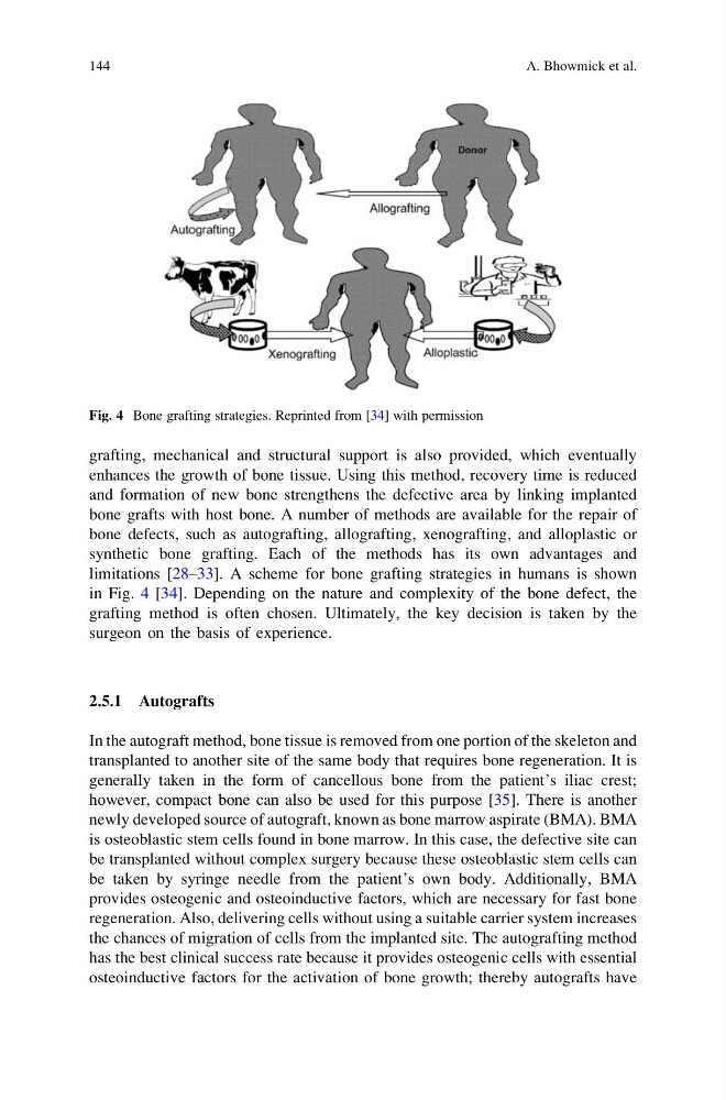
limitations [28–33]. A scheme for bone grafting strategies in humans is shown
in Fig. 4 [34]. Depending on the nature and complexity of the bone defect, the
grafting method is often chosen. Ultimately, the key decision is taken by the
surgeon on the basis of experience.
2.5.1 Autografts
In the autograft method, bone tissue is removed from one portion of the skeleton and
transplanted to another site of the same body that requires bone regeneration. It is
generally taken in the form of cancellous bone from the patient’s iliac crest;
however, compact bone can also be used for this purpose [35]. There is another
newly developed source of autograft, known as bone marrow aspirate (BMA). BMA
is osteoblastic stem cells found in bone marrow. In this case, the defective site can
be transplanted without complex surgery because these osteoblastic stem cells can
be taken by syringe needle from the patient’s own body. Additionally, BMA
provides osteogenic and osteoinductive factors, which are necessary for fast bone
regeneration. Also, delivering cells without using a suitable carrier system increases
the chances of migration of cells from the implanted site. The autografting method
has the best clinical success rate because it provides osteogenic cells with essential
osteoinductive factors for the activation of bone growth; thereby autografts have
Fig. 4 Bone grafting strategies. Reprinted from [34] with permission
144
A. Bhowmick et al.
enhances the growth of bone tissue. Using this method, recovery time is reduced
and formation of new bone strengthens the defective area by linking implanted
bone grafts with host bone. A number of methods are available for the repair of
bone defects, such as autografting, allografting, xenografting, and alloplastic or
synthetic bone grafting. Each of the methods has its own advantages and
limitations [28–33]. A scheme for bone grafting strategies in humans is shown
in Fig. 4 [34]. Depending on the nature and complexity of the bone defect, the
grafting method is often chosen. Ultimately, the key decision is taken by the
surgeon on the basis of experience.
2.5.1 Autografts
In the autograft method, bone tissue is removed from one portion of the skeleton and
transplanted to another site of the same body that requires bone regeneration. It is
generally taken in the form of cancellous bone from the patient’s iliac crest;
however, compact bone can also be used for this purpose [35]. There is another
newly developed source of autograft, known as bone marrow aspirate (BMA). BMA
is osteoblastic stem cells found in bone marrow. In this case, the defective site can
be transplanted without complex surgery because these osteoblastic stem cells can
be taken by syringe needle from the patient’s own body. Additionally, BMA
provides osteogenic and osteoinductive factors, which are necessary for fast bone
regeneration. Also, delivering cells without using a suitable carrier system increases
the chances of migration of cells from the implanted site. The autografting method
has the best clinical success rate because it provides osteogenic cells with essential
osteoinductive factors for the activation of bone growth; thereby autografts have
Fig. 4 Bone grafting strategies. Reprinted from [34] with permission
144
A. Bhowmick et al.