

investigated. Both blocks possess helices stabilized either by hydrogen bonds (PBLG)
or by steric hindrance (PLP) and are packed in two hexagonal cells of different
dimensions. An intriguing trans–cis conformational change of PLP upon confinement
was observed that mimics the isomerization of isolated proline residues in proteins.
These cis-PLP conformations reside primarily at the PLP/PBLG interface, alleviate
the packing frustration (see Fig. 4), and permit PBLG and PLP helices to pack with
the bulk [59].
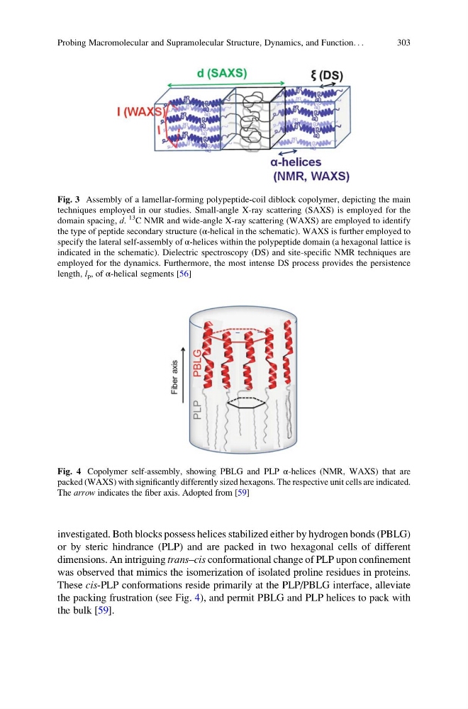
Fig. 3 Assembly of a lamellar-forming polypeptide-coil diblock copolymer, depicting the main
techniques employed in our studies. Small-angle X-ray scattering (SAXS) is employed for the
domain spacing, d.
13
C NMR and wide-angle X-ray scattering (WAXS) are employed to identify
the type of peptide secondary structure (α-helical in the schematic). WAXS is further employed to
specify the lateral self-assembly of α-helices within the polypeptide domain (a hexagonal lattice is
indicated in the schematic). Dielectric spectroscopy (DS) and site-specific NMR techniques are
employed for the dynamics. Furthermore, the most intense DS process provides the persistence
length, l p , of α-helical segments [56]
Fig. 4 Copolymer self-assembly, showing PBLG and PLP α-helices (NMR, WAXS) that are
packed (WAXS) with significantly differently sized hexagons. The respective unit cells are indicated.
The arrow indicates the fiber axis. Adopted from [59]
Probing Macromolecular and Supramolecular Structure, Dynamics, and Function. . .
303
or by steric hindrance (PLP) and are packed in two hexagonal cells of different
dimensions. An intriguing trans–cis conformational change of PLP upon confinement
was observed that mimics the isomerization of isolated proline residues in proteins.
These cis-PLP conformations reside primarily at the PLP/PBLG interface, alleviate
the packing frustration (see Fig. 4), and permit PBLG and PLP helices to pack with
the bulk [59].
Fig. 3 Assembly of a lamellar-forming polypeptide-coil diblock copolymer, depicting the main
techniques employed in our studies. Small-angle X-ray scattering (SAXS) is employed for the
domain spacing, d.
13
C NMR and wide-angle X-ray scattering (WAXS) are employed to identify
the type of peptide secondary structure (α-helical in the schematic). WAXS is further employed to
specify the lateral self-assembly of α-helices within the polypeptide domain (a hexagonal lattice is
indicated in the schematic). Dielectric spectroscopy (DS) and site-specific NMR techniques are
employed for the dynamics. Furthermore, the most intense DS process provides the persistence
length, l p , of α-helical segments [56]
Fig. 4 Copolymer self-assembly, showing PBLG and PLP α-helices (NMR, WAXS) that are
packed (WAXS) with significantly differently sized hexagons. The respective unit cells are indicated.
The arrow indicates the fiber axis. Adopted from [59]
Probing Macromolecular and Supramolecular Structure, Dynamics, and Function. . .
303