

5 Conclusions
In this chapter we have presented an overview of different approaches to the study
of lipid membranes and membrane protein systems. We have reviewed theoretical
and simulation approaches, and shown how generic lipid simulation models can be
used to understand the principles that determine properties of lipid bilayers such as
bending, Gaussian curvature modulus, and membrane tension, or fundamental
phenomena such as the formation of lipid rafts or the curvature-mediated interactions between proteins. In the previous section it was outlined how multiscale
modeling can in principle go a step further by ensuring a certain chemical specificity while still benefiting from the time- and length-scale advantages of coarsegrained simulations. It was noted that there are still a number of challenges in the
area of systematic coarse graining that need to be addressed to be able to study
complex multicomponent systems such as the light-harvesting complex of green
plants. For this system, we have shown the first steps toward a multiscale simulation
model that allows going back and forth between a coarse-grained and an atomistic
level of resolution and therefore permits immediate comparison to atomic level
experimental data.
Acknowledgements We would like to thank the many coworkers and colleagues who have
contributed to the research reported here, in particular Ira Cooke, Jemal Guven, Vagelis
Harmandaris, Gregoria Illya, Martin Mu ¨ller, Benedict Reynwar, Ira Rothstein, Cem Yolcu,
Frank Brown, Olaf Lenz, Sebastian Meinhardt, Peter Nielaba, Beate West, Ananya Debnath,
Christoph Globisch, Christoph Junghans, Shahoua Ding, Sabine Wiegand, Sandra Ritz, and Eva
Sinner.
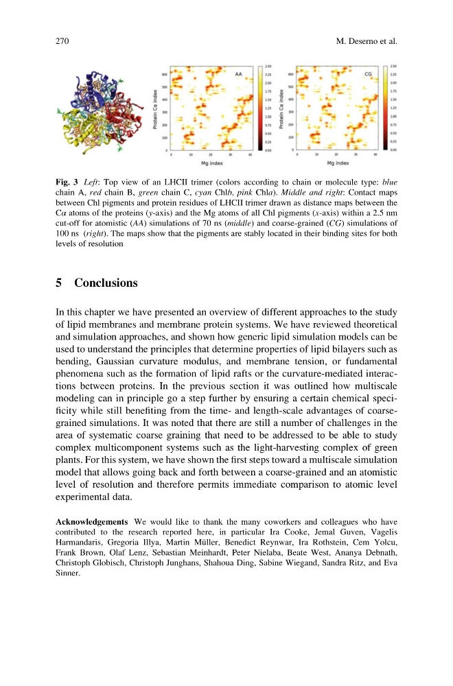
Fig. 3 Left: Top view of an LHCII trimer (colors according to chain or molecule type: blue
chain A, red chain B, green chain C, cyan Chlb, pink Chla). Middle and right: Contact maps
between Chl pigments and protein residues of LHCII trimer drawn as distance maps between the
Cα atoms of the proteins (y-axis) and the Mg atoms of all Chl pigments (x-axis) within a 2.5 nm
cut-off for atomistic (AA) simulations of 70 ns (middle) and coarse-grained (CG) simulations of
100 ns (right). The maps show that the pigments are stably located in their binding sites for both
levels of resolution
270
M. Deserno et al.
In this chapter we have presented an overview of different approaches to the study
of lipid membranes and membrane protein systems. We have reviewed theoretical
and simulation approaches, and shown how generic lipid simulation models can be
used to understand the principles that determine properties of lipid bilayers such as
bending, Gaussian curvature modulus, and membrane tension, or fundamental
phenomena such as the formation of lipid rafts or the curvature-mediated interactions between proteins. In the previous section it was outlined how multiscale
modeling can in principle go a step further by ensuring a certain chemical specificity while still benefiting from the time- and length-scale advantages of coarsegrained simulations. It was noted that there are still a number of challenges in the
area of systematic coarse graining that need to be addressed to be able to study
complex multicomponent systems such as the light-harvesting complex of green
plants. For this system, we have shown the first steps toward a multiscale simulation
model that allows going back and forth between a coarse-grained and an atomistic
level of resolution and therefore permits immediate comparison to atomic level
experimental data.
Acknowledgements We would like to thank the many coworkers and colleagues who have
contributed to the research reported here, in particular Ira Cooke, Jemal Guven, Vagelis
Harmandaris, Gregoria Illya, Martin Mu ¨ller, Benedict Reynwar, Ira Rothstein, Cem Yolcu,
Frank Brown, Olaf Lenz, Sebastian Meinhardt, Peter Nielaba, Beate West, Ananya Debnath,
Christoph Globisch, Christoph Junghans, Shahoua Ding, Sabine Wiegand, Sandra Ritz, and Eva
Sinner.
Fig. 3 Left: Top view of an LHCII trimer (colors according to chain or molecule type: blue
chain A, red chain B, green chain C, cyan Chlb, pink Chla). Middle and right: Contact maps
between Chl pigments and protein residues of LHCII trimer drawn as distance maps between the
Cα atoms of the proteins (y-axis) and the Mg atoms of all Chl pigments (x-axis) within a 2.5 nm
cut-off for atomistic (AA) simulations of 70 ns (middle) and coarse-grained (CG) simulations of
100 ns (right). The maps show that the pigments are stably located in their binding sites for both
levels of resolution
270
M. Deserno et al.