

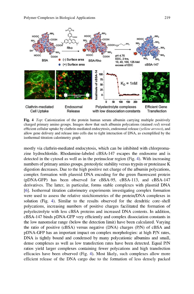
mostly via clathrin-mediated endocytosis, which can be inhibited with chlorpromazine hydrochloride. Rhodamine-labeled cBSA-147 escapes the endosome and is
detected in the cytosol as well as in the perinuclear region (Fig. 4). With increasing
numbers of primary amino groups, proteolytic stability versus trypsin or proteinase K
digestion decreases. Due to the high positive net charge of the albumin polycations,
complex formation with plasmid DNA encoding for the green fluorescent protein
(pDNA-GFP) has been observed for cBSA-95, cBSA-113, and cBSA-147
derivatives. The latter, in particular, forms stable complexes with plasmid DNA
[6]. Isothermal titration calorimetry experiments investigating complex formation
were used to assess the relative stoichiometries of the protein/DNA complexes in
solution (Fig. 4). Similar to the results observed for the dendritic core–shell
polycations, increasing numbers of positive charges facilitated the formation of
polyelectrolyte with less cBSA proteins and increased DNA contents. In addition,
cBSA-147 binds pDNA-GFP very efficiently and complex dissociation constants in
the low nanomolar range (below the detection limit) have been calculated. Varying
the ratio of positive (cBSA) versus negative (DNA) charges (P/N) of cBSA and
pDNA-GFP has an important impact on complex morphologies: at high P/N rates,
DNA is tightly bound and condensed by many polycationic albumins and small,
dense complexes as well as low transfection rates have been detected. Equal P/N
ratios yield larger complexes containing fewer polycations and high transfection
efficacies have been observed (Fig. 4). Most likely, such complexes allow more
efficient release of the DNA cargo due to the formation of less densely packed
Fig. 4 Top: Cationization of the protein human serum albumin carrying multiple positively
charged primary amino groups. Images show that such albumin polycations (stained red) reveal
efficient cellular uptake by clathrin-mediated endocytosis, endosomal release (yellow arrows), and
allow gene delivery and release into cells due to tight interaction of DNA, as exemplified by the
isothermal titration calorimetry graph
Polymer Complexes in Biological Applications
219
detected in the cytosol as well as in the perinuclear region (Fig. 4). With increasing
numbers of primary amino groups, proteolytic stability versus trypsin or proteinase K
digestion decreases. Due to the high positive net charge of the albumin polycations,
complex formation with plasmid DNA encoding for the green fluorescent protein
(pDNA-GFP) has been observed for cBSA-95, cBSA-113, and cBSA-147
derivatives. The latter, in particular, forms stable complexes with plasmid DNA
[6]. Isothermal titration calorimetry experiments investigating complex formation
were used to assess the relative stoichiometries of the protein/DNA complexes in
solution (Fig. 4). Similar to the results observed for the dendritic core–shell
polycations, increasing numbers of positive charges facilitated the formation of
polyelectrolyte with less cBSA proteins and increased DNA contents. In addition,
cBSA-147 binds pDNA-GFP very efficiently and complex dissociation constants in
the low nanomolar range (below the detection limit) have been calculated. Varying
the ratio of positive (cBSA) versus negative (DNA) charges (P/N) of cBSA and
pDNA-GFP has an important impact on complex morphologies: at high P/N rates,
DNA is tightly bound and condensed by many polycationic albumins and small,
dense complexes as well as low transfection rates have been detected. Equal P/N
ratios yield larger complexes containing fewer polycations and high transfection
efficacies have been observed (Fig. 4). Most likely, such complexes allow more
efficient release of the DNA cargo due to the formation of less densely packed
Fig. 4 Top: Cationization of the protein human serum albumin carrying multiple positively
charged primary amino groups. Images show that such albumin polycations (stained red) reveal
efficient cellular uptake by clathrin-mediated endocytosis, endosomal release (yellow arrows), and
allow gene delivery and release into cells due to tight interaction of DNA, as exemplified by the
isothermal titration calorimetry graph
Polymer Complexes in Biological Applications
219