

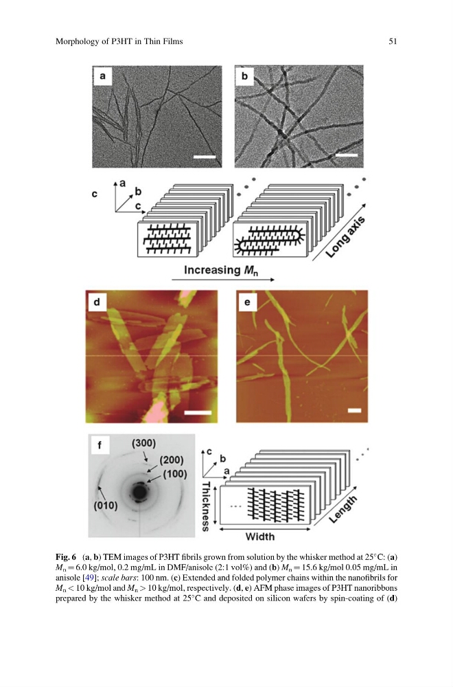
Fig. 6 (a, b) TEM images of P3HT fibrils grown from solution by the whisker method at 25
C: (a)
M n ¼ 6.0 kg/mol, 0.2 mg/mL in DMF/anisole (2:1 vol%) and (b) M n ¼ 15.6 kg/mol 0.05 mg/mL in
anisole [49]; scale bars: 100 nm. (c) Extended and folded polymer chains within the nanofibrils for
M n < 10 kg/mol and M n > 10 kg/mol, respectively. (d, e) AFM phase images of P3HT nanoribbons
prepared by the whisker method at 25
C and deposited on silicon wafers by spin-coating of (d)
Morphology of P3HT in Thin Films
51
C: (a)
M n ¼ 6.0 kg/mol, 0.2 mg/mL in DMF/anisole (2:1 vol%) and (b) M n ¼ 15.6 kg/mol 0.05 mg/mL in
anisole [49]; scale bars: 100 nm. (c) Extended and folded polymer chains within the nanofibrils for
M n < 10 kg/mol and M n > 10 kg/mol, respectively. (d, e) AFM phase images of P3HT nanoribbons
prepared by the whisker method at 25
C and deposited on silicon wafers by spin-coating of (d)
Morphology of P3HT in Thin Films
51