

proteins, reducing the pH of the protein solution subjected to heat treatment speeds up
the formation of fibrils regardless of the pI of native protein, for instance LYS (10.7)
and α-La (4.2–4.5). Heat treatment under acidic pH conditions induces some cleavage in the polypeptide backbone [76, 82, 85] and generates some succinimidyl
residues from aspartic acid [76], both reactions being preponderant at low pH.
Peptides are considered to be the major building blocks of fibrils, but the contribution
of succinimdyl residues is much more speculative. For β-Lg, the yield of
incorporation of the peptides into fibrils is low because only the most hydrophobic
peptides with β-sheet propensity are involved [85]. The shape and size of the fibrils
are also tuned by ionic strength. Screening of the electrostatic charges by salts fastens
the aggregation stage, resulting in shorter and more curved fibrils [89]. However,
according to these authors, the fibrils exhibited similar thicknesses of around 3.5 nm
and a periodic structure, with a period of about 25 nm whatever the ionic strength in
the range 10–100 mM.
3.2 Multistranded Ribbons and Spherulites
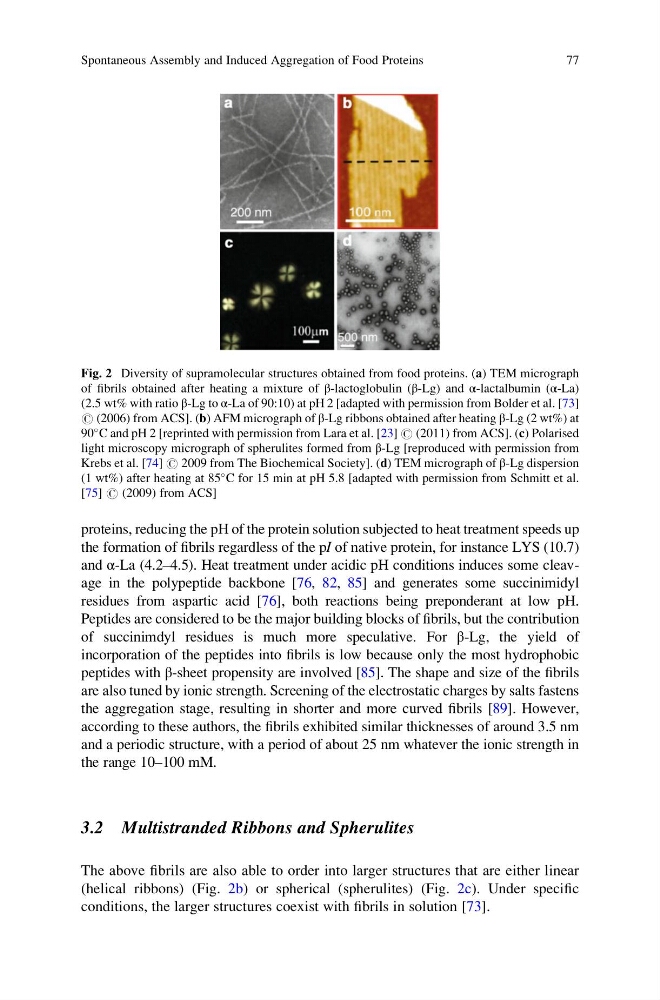
The above fibrils are also able to order into larger structures that are either linear
(helical ribbons) (Fig. 2b) or spherical (spherulites) (Fig. 2c). Under specific
conditions, the larger structures coexist with fibrils in solution [73].
Fig. 2 Diversity of supramolecular structures obtained from food proteins. (a) TEM micrograph
of fibrils obtained after heating a mixture of β-lactoglobulin (β-Lg) and α-lactalbumin (α-La)
(2.5 wt% with ratio β-Lg to α-La of 90:10) at pH 2 [adapted with permission from Bolder et al. [73]
# (2006) from ACS]. (b) AFM micrograph of β-Lg ribbons obtained after heating β-Lg (2 wt%) at
90
C and pH 2 [reprinted with permission from Lara et al. [23] # (2011) from ACS]. (c) Polarised
light microscopy micrograph of spherulites formed from β-Lg [reproduced with permission from
Krebs et al. [74] # 2009 from The Biochemical Society]. (d) TEM micrograph of β-Lg dispersion
(1 wt%) after heating at 85
C for 15 min at pH 5.8 [adapted with permission from Schmitt et al.
[75] # (2009) from ACS]
Spontaneous Assembly and Induced Aggregation of Food Proteins
77
the formation of fibrils regardless of the pI of native protein, for instance LYS (10.7)
and α-La (4.2–4.5). Heat treatment under acidic pH conditions induces some cleavage in the polypeptide backbone [76, 82, 85] and generates some succinimidyl
residues from aspartic acid [76], both reactions being preponderant at low pH.
Peptides are considered to be the major building blocks of fibrils, but the contribution
of succinimdyl residues is much more speculative. For β-Lg, the yield of
incorporation of the peptides into fibrils is low because only the most hydrophobic
peptides with β-sheet propensity are involved [85]. The shape and size of the fibrils
are also tuned by ionic strength. Screening of the electrostatic charges by salts fastens
the aggregation stage, resulting in shorter and more curved fibrils [89]. However,
according to these authors, the fibrils exhibited similar thicknesses of around 3.5 nm
and a periodic structure, with a period of about 25 nm whatever the ionic strength in
the range 10–100 mM.
3.2 Multistranded Ribbons and Spherulites
The above fibrils are also able to order into larger structures that are either linear
(helical ribbons) (Fig. 2b) or spherical (spherulites) (Fig. 2c). Under specific
conditions, the larger structures coexist with fibrils in solution [73].
Fig. 2 Diversity of supramolecular structures obtained from food proteins. (a) TEM micrograph
of fibrils obtained after heating a mixture of β-lactoglobulin (β-Lg) and α-lactalbumin (α-La)
(2.5 wt% with ratio β-Lg to α-La of 90:10) at pH 2 [adapted with permission from Bolder et al. [73]
# (2006) from ACS]. (b) AFM micrograph of β-Lg ribbons obtained after heating β-Lg (2 wt%) at
90
C and pH 2 [reprinted with permission from Lara et al. [23] # (2011) from ACS]. (c) Polarised
light microscopy micrograph of spherulites formed from β-Lg [reproduced with permission from
Krebs et al. [74] # 2009 from The Biochemical Society]. (d) TEM micrograph of β-Lg dispersion
(1 wt%) after heating at 85
C for 15 min at pH 5.8 [adapted with permission from Schmitt et al.
[75] # (2009) from ACS]
Spontaneous Assembly and Induced Aggregation of Food Proteins
77