

Apart from these two references, we could not find further open literature reports on
the interaction between PEC nanoparticles and endothelial cells.
However, the fact that PEC material positively interacts with HUVECs was
noticed earlier by Boura et al. using the related PEM of PAH/PSS or PLL/PLG
[171]. It was shown that these PEM enhanced initial cell adhesion compared with
single PEL layers and are neither cytotoxic nor change the HUVEC phenotype.
Recently, related collagen/heparin multilayers were shown to have similar positive
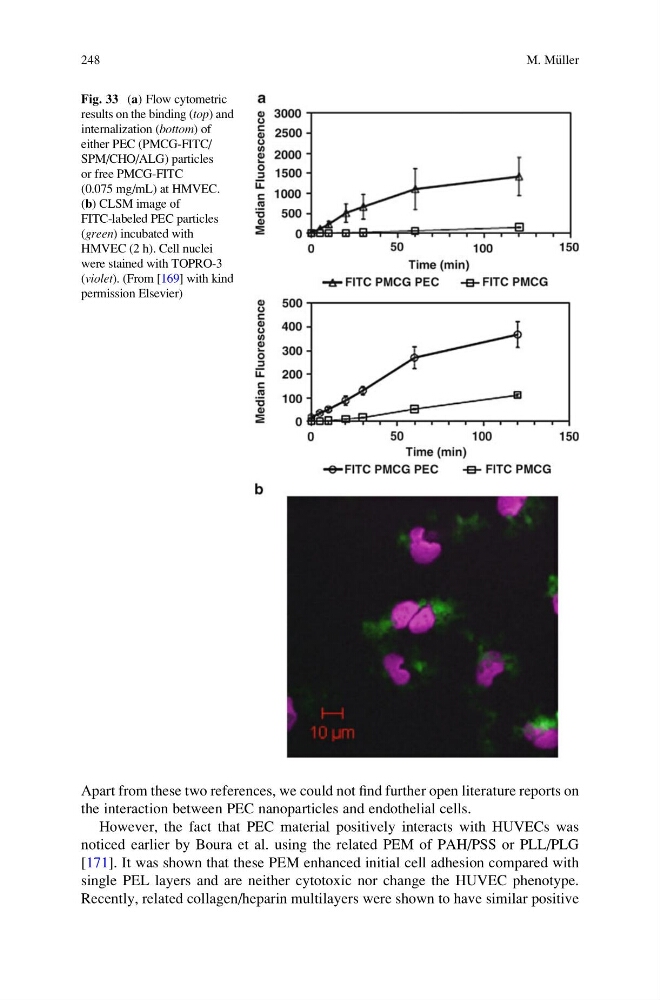
Fig. 33 (a) Flow cytometric
results on the binding (top) and
internalization (bottom) of
either PEC (PMCG-FITC/
SPM/CHO/ALG) particles
or free PMCG-FITC
(0.075 mg/mL) at HMVEC.
(b) CLSM image of
FITC-labeled PEC particles
(green) incubated with
HMVEC (2 h). Cell nuclei
were stained with TOPRO-3
(violet). (From [169] with kind
permission Elsevier)
248
M. M€ uller
the interaction between PEC nanoparticles and endothelial cells.
However, the fact that PEC material positively interacts with HUVECs was
noticed earlier by Boura et al. using the related PEM of PAH/PSS or PLL/PLG
[171]. It was shown that these PEM enhanced initial cell adhesion compared with
single PEL layers and are neither cytotoxic nor change the HUVEC phenotype.
Recently, related collagen/heparin multilayers were shown to have similar positive
Fig. 33 (a) Flow cytometric
results on the binding (top) and
internalization (bottom) of
either PEC (PMCG-FITC/
SPM/CHO/ALG) particles
or free PMCG-FITC
(0.075 mg/mL) at HMVEC.
(b) CLSM image of
FITC-labeled PEC particles
(green) incubated with
HMVEC (2 h). Cell nuclei
were stained with TOPRO-3
(violet). (From [169] with kind
permission Elsevier)
248
M. M€ uller