

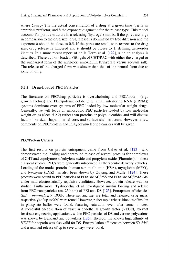
where C DRUG (t) is the actual concentration of a drug at a given time t, a is an
empirical prefactor, and b the exponent diagnostic for the release type. This model
accounts for porous structure in a releasing (hydrogel) matrix. If the pores are large
in comparison to the drug size, drug release is dominated by free diffusion and the
exponent b should be close to 0.5. If the pores are small with respect to the drug
size, drug release is hindered and b should be closer to 1, defining zero-order
kinetics. In a more recent report of de la Torre et al. [122], such an analysis is
described. These authors loaded PEC gels of CHT/PAC with either the charged or
the uncharged form of the antibiotic amoxicillin (trihydrate versus sodium salt).
The release of the charged form was slower than that of the neutral form due to
ionic binding.
5.2.2 Drug-Loaded PEC Particles
The literature on PEC/drug particles is overwhelming and PEC/protein (e.g.,
growth factors) and PEC/polynucleotide (e.g., small interfering RNA (siRNA))
systems dominate over systems of PEC loaded by low molecular weight drugs.
Generally, we will focus on nanoscopic PEC particles loaded by low molecular
weight drugs (Sect. 5.2.2) rather than proteins or polynucleotides and will discuss
factors like size, shape, internal core, and surface shell structure. However, a few
comments on PEC/protein and PEC/polynucleotide carriers will be given.
PEC/Protein Carriers
The first results on protein entrapment came from Calvo et al. [123], who
demonstrated the loading and controlled release of several proteins for complexes
of CHT and copolymers of ethylene oxide and propylene oxide (Pluronics). In these
classical studies, PECs were generally introduced as therapeutic delivery vehicles.
Loading of the model proteins human serum albumin (HSA), myoglobin (MYO),
and lysozyme (LYZ) has also been shown by Ouyang and M€ uller [124]. These
proteins were bound to PEC particles of PDADMAC/PSS and PDADMAC/PMA-MS
under mild electrostatically repulsive conditions. However, protein release was not
studied. Furthermore, Tyaboonchai et al. investigated insulin loading and release
from PEC nanoparticles (ca. 250 nm) of PEI and DS [125]. Entrapment efficiencies
(EE ¼ m 0 Àm R /m 0 Â 100%, where m 0 and m R are total and released drug mass,
respectively) of up to 90% were found. However, rather rapid release kinetics of insulin
in phosphate buffer were found, featuring saturation even after some minutes.
A successful encapsulation of vascular endothelial growth factor (VEGF), relevant
for tissue engineering applications, within PEC particles of DS and various polycations
was shown by Berkland and coworkers [126]. Thereby, the known high affinity of
VEGF for heparin was also valid for DS. Encapsulation efficiencies between 50–85%
and a retarded release of up to several days were found.
Sizing, Shaping and Pharmaceutical Applications of Polyelectrolyte Complex. . .
237
empirical prefactor, and b the exponent diagnostic for the release type. This model
accounts for porous structure in a releasing (hydrogel) matrix. If the pores are large
in comparison to the drug size, drug release is dominated by free diffusion and the
exponent b should be close to 0.5. If the pores are small with respect to the drug
size, drug release is hindered and b should be closer to 1, defining zero-order
kinetics. In a more recent report of de la Torre et al. [122], such an analysis is
described. These authors loaded PEC gels of CHT/PAC with either the charged or
the uncharged form of the antibiotic amoxicillin (trihydrate versus sodium salt).
The release of the charged form was slower than that of the neutral form due to
ionic binding.
5.2.2 Drug-Loaded PEC Particles
The literature on PEC/drug particles is overwhelming and PEC/protein (e.g.,
growth factors) and PEC/polynucleotide (e.g., small interfering RNA (siRNA))
systems dominate over systems of PEC loaded by low molecular weight drugs.
Generally, we will focus on nanoscopic PEC particles loaded by low molecular
weight drugs (Sect. 5.2.2) rather than proteins or polynucleotides and will discuss
factors like size, shape, internal core, and surface shell structure. However, a few
comments on PEC/protein and PEC/polynucleotide carriers will be given.
PEC/Protein Carriers
The first results on protein entrapment came from Calvo et al. [123], who
demonstrated the loading and controlled release of several proteins for complexes
of CHT and copolymers of ethylene oxide and propylene oxide (Pluronics). In these
classical studies, PECs were generally introduced as therapeutic delivery vehicles.
Loading of the model proteins human serum albumin (HSA), myoglobin (MYO),
and lysozyme (LYZ) has also been shown by Ouyang and M€ uller [124]. These
proteins were bound to PEC particles of PDADMAC/PSS and PDADMAC/PMA-MS
under mild electrostatically repulsive conditions. However, protein release was not
studied. Furthermore, Tyaboonchai et al. investigated insulin loading and release
from PEC nanoparticles (ca. 250 nm) of PEI and DS [125]. Entrapment efficiencies
(EE ¼ m 0 Àm R /m 0 Â 100%, where m 0 and m R are total and released drug mass,
respectively) of up to 90% were found. However, rather rapid release kinetics of insulin
in phosphate buffer were found, featuring saturation even after some minutes.
A successful encapsulation of vascular endothelial growth factor (VEGF), relevant
for tissue engineering applications, within PEC particles of DS and various polycations
was shown by Berkland and coworkers [126]. Thereby, the known high affinity of
VEGF for heparin was also valid for DS. Encapsulation efficiencies between 50–85%
and a retarded release of up to several days were found.
Sizing, Shaping and Pharmaceutical Applications of Polyelectrolyte Complex. . .
237