

52
3 Materials and Methods
Gram Staining Procedure of Bacteria
1. One clean slide was gently heated in the flame to remove any grease present.
2. A loopful of distilled water was placed in the center of the slide.
3. A minute amount of bacterial growth with the help of a wire loop was removed.
4. The bacterial growth was emulsified in a loopful of distilled water on the slide.
The smear was prepared by spreading the suspension in a thin film on the slide.
5. The smear was fixed quickly passing the slide through the flame.
6. The film was covered with sufficient crystal violet stain and allowed to remain
for 1 min.
7. The excess stain was passed off and washed the slide with tap water.
8. The film was covered with Gram’s iodine solution and was allowed to stand for
1 min.
9. The slide was washed on tap water and dried.
10. 95% of ethyl alcohol was passed dropwise till the violet color flowed away.
11. The slide was covered with Safranin for 30 s.
12. The slide was washed in tap water and blotted dry.
13. Then, the slide was examined under microscope.
3.9.1.6 Bacterial Characterization Using 16S rDNA Protocol
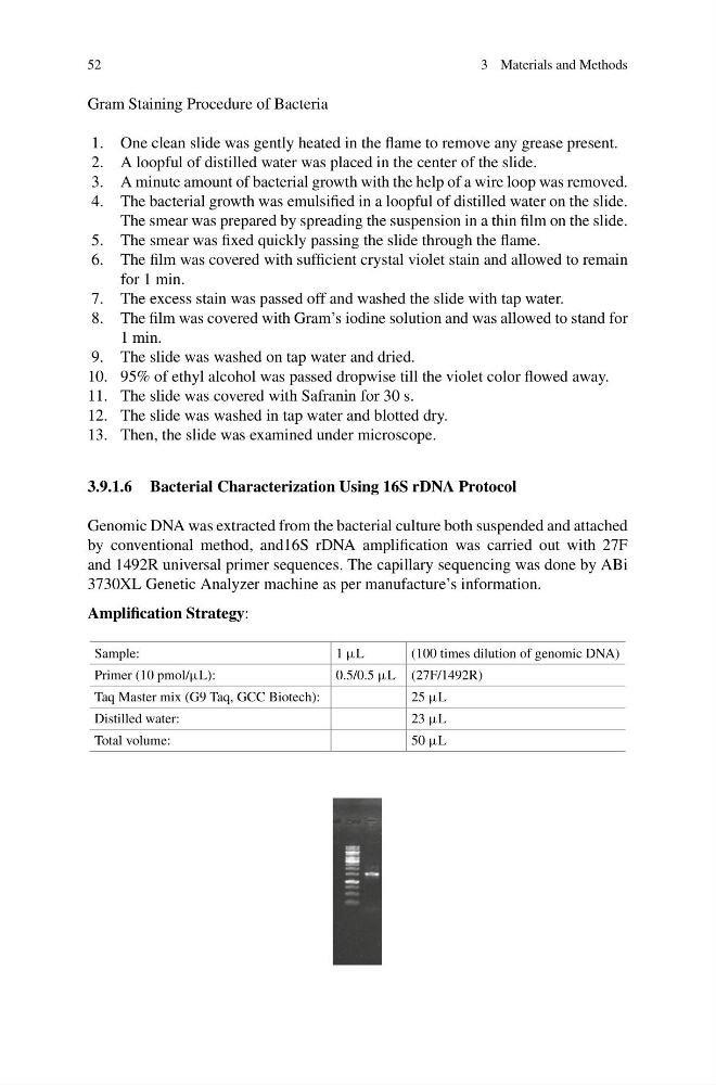
Genomic DNA was extracted from the bacterial culture both suspended and attached
by conventional method, and16S rDNA amplification was carried out with 27F
and 1492R universal primer sequences. The capillary sequencing was done by ABi
3730XL Genetic Analyzer machine as per manufacture’s information.
Amplification Strategy:
Sample:
1 µL
(100 times dilution of genomic DNA)
Primer (10 pmol/µL):
0.5/0.5 µL (27F/1492R)
Taq Master mix (G9 Taq, GCC Biotech):
25 µL
Distilled water:
23 µL
Total volume:
50 µL
3 Materials and Methods
Gram Staining Procedure of Bacteria
1. One clean slide was gently heated in the flame to remove any grease present.
2. A loopful of distilled water was placed in the center of the slide.
3. A minute amount of bacterial growth with the help of a wire loop was removed.
4. The bacterial growth was emulsified in a loopful of distilled water on the slide.
The smear was prepared by spreading the suspension in a thin film on the slide.
5. The smear was fixed quickly passing the slide through the flame.
6. The film was covered with sufficient crystal violet stain and allowed to remain
for 1 min.
7. The excess stain was passed off and washed the slide with tap water.
8. The film was covered with Gram’s iodine solution and was allowed to stand for
1 min.
9. The slide was washed on tap water and dried.
10. 95% of ethyl alcohol was passed dropwise till the violet color flowed away.
11. The slide was covered with Safranin for 30 s.
12. The slide was washed in tap water and blotted dry.
13. Then, the slide was examined under microscope.
3.9.1.6 Bacterial Characterization Using 16S rDNA Protocol
Genomic DNA was extracted from the bacterial culture both suspended and attached
by conventional method, and16S rDNA amplification was carried out with 27F
and 1492R universal primer sequences. The capillary sequencing was done by ABi
3730XL Genetic Analyzer machine as per manufacture’s information.
Amplification Strategy:
Sample:
1 µL
(100 times dilution of genomic DNA)
Primer (10 pmol/µL):
0.5/0.5 µL (27F/1492R)
Taq Master mix (G9 Taq, GCC Biotech):
25 µL
Distilled water:
23 µL
Total volume:
50 µL