

268
S. E. H. Murph and E. Searles
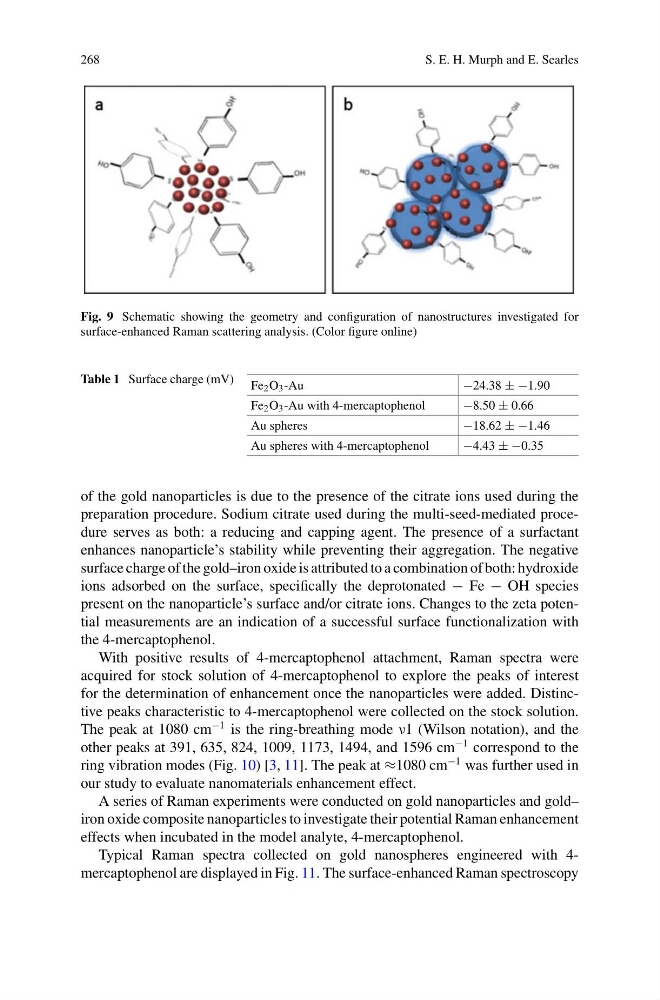
Fig. 9 Schematic showing the geometry and configuration of nanostructures investigated for
surface-enhanced Raman scattering analysis. (Color figure online)
Table 1 Surface charge (mV) Fe 2 O 3 -Au
−24.38 ± −1.90
Fe 2 O 3 -Au with 4-mercaptophenol
−8.50 ± 0.66
Au spheres
−18.62 ± −1.46
Au spheres with 4-mercaptophenol
−4.43 ± −0.35
of the gold nanoparticles is due to the presence of the citrate ions used during the
preparation procedure. Sodium citrate used during the multi-seed-mediated procedure serves as both: a reducing and capping agent. The presence of a surfactant
enhances nanoparticle’s stability while preventing their aggregation. The negative
surface charge of the gold–iron oxide is attributed to a combination of both: hydroxide
ions adsorbed on the surface, specifically the deprotonated − Fe − OH species
present on the nanoparticle’s surface and/or citrate ions. Changes to the zeta potential measurements are an indication of a successful surface functionalization with
the 4-mercaptophenol.
With positive results of 4-mercaptophenol attachment, Raman spectra were
acquired for stock solution of 4-mercaptophenol to explore the peaks of interest
for the determination of enhancement once the nanoparticles were added. Distinctive peaks characteristic to 4-mercaptophenol were collected on the stock solution.
The peak at 1080 cm
−1 is the ring-breathing mode ν1 (Wilson notation), and the
other peaks at 391, 635, 824, 1009, 1173, 1494, and 1596 cm
−1 correspond to the
ring vibration modes (Fig. 10) [3, 11]. The peak at ≈1080 cm
−1 was further used in
our study to evaluate nanomaterials enhancement effect.
A series of Raman experiments were conducted on gold nanoparticles and gold–
iron oxide composite nanoparticles to investigate their potential Raman enhancement
effects when incubated in the model analyte, 4-mercaptophenol.
Typical Raman spectra collected on gold nanospheres engineered with 4mercaptophenol are displayed in Fig. 11. The surface-enhanced Raman spectroscopy
S. E. H. Murph and E. Searles
Fig. 9 Schematic showing the geometry and configuration of nanostructures investigated for
surface-enhanced Raman scattering analysis. (Color figure online)
Table 1 Surface charge (mV) Fe 2 O 3 -Au
−24.38 ± −1.90
Fe 2 O 3 -Au with 4-mercaptophenol
−8.50 ± 0.66
Au spheres
−18.62 ± −1.46
Au spheres with 4-mercaptophenol
−4.43 ± −0.35
of the gold nanoparticles is due to the presence of the citrate ions used during the
preparation procedure. Sodium citrate used during the multi-seed-mediated procedure serves as both: a reducing and capping agent. The presence of a surfactant
enhances nanoparticle’s stability while preventing their aggregation. The negative
surface charge of the gold–iron oxide is attributed to a combination of both: hydroxide
ions adsorbed on the surface, specifically the deprotonated − Fe − OH species
present on the nanoparticle’s surface and/or citrate ions. Changes to the zeta potential measurements are an indication of a successful surface functionalization with
the 4-mercaptophenol.
With positive results of 4-mercaptophenol attachment, Raman spectra were
acquired for stock solution of 4-mercaptophenol to explore the peaks of interest
for the determination of enhancement once the nanoparticles were added. Distinctive peaks characteristic to 4-mercaptophenol were collected on the stock solution.
The peak at 1080 cm
−1 is the ring-breathing mode ν1 (Wilson notation), and the
other peaks at 391, 635, 824, 1009, 1173, 1494, and 1596 cm
−1 correspond to the
ring vibration modes (Fig. 10) [3, 11]. The peak at ≈1080 cm
−1 was further used in
our study to evaluate nanomaterials enhancement effect.
A series of Raman experiments were conducted on gold nanoparticles and gold–
iron oxide composite nanoparticles to investigate their potential Raman enhancement
effects when incubated in the model analyte, 4-mercaptophenol.
Typical Raman spectra collected on gold nanospheres engineered with 4mercaptophenol are displayed in Fig. 11. The surface-enhanced Raman spectroscopy