

11.4 Soft Templates Formed by Molecular Self-assembly Processes
403
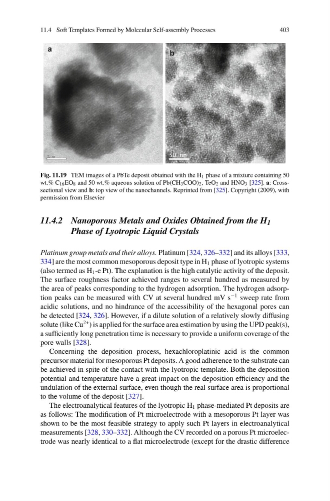
Fig. 11.19 TEM images of a PbTe deposit obtained with the H 1 phase of a mixture containing 50
wt.% C 16 EO 8 and 50 wt.% aqueous solution of Pb(CH 3 COO) 2 , TeO 2 and HNO 3 [325]. a: Crosssectional view and b: top view of the nanochannels. Reprinted from [325]. Copyright (2009), with
permission from Elsevier
11.4.2 Nanoporous Metals and Oxides Obtained from the H 1
Phase of Lyotropic Liquid Crystals
Platinum group metals and their alloys. Platinum [324, 326–332] and its alloys [333,
334] are the most common mesoporous deposit type in H 1 phase of lyotropic systems
(also termed as H 1 -e Pt). The explanation is the high catalytic activity of the deposit.
The surface roughness factor achieved ranges to several hundred as measured by
the area of peaks corresponding to the hydrogen adsorption. The hydrogen adsorption peaks can be measured with CV at several hundred mV s
−1 sweep rate from
acidic solutions, and no hindrance of the accessibility of the hexagonal pores can
be detected [324, 326]. However, if a dilute solution of a relatively slowly diffusing
solute (like Cu
2+ ) is applied for the surface area estimation by using the UPD peak(s),
a sufficiently long penetration time is necessary to provide a uniform coverage of the
pore walls [328].
Concerning the deposition process, hexachloroplatinic acid is the common
precursor material for mesoporous Pt deposits. A good adherence to the substrate can
be achieved in spite of the contact with the lyotropic template. Both the deposition
potential and temperature have a great impact on the deposition efficiency and the
undulation of the external surface, even though the real surface area is proportional
to the volume of the deposit [327].
The electroanalytical features of the lyotropic H 1 phase-mediated Pt deposits are
as follows: The modification of Pt microelectrode with a mesoporous Pt layer was
shown to be the most feasible strategy to apply such Pt layers in electroanalytical
measurements [328, 330–332]. Although the CV recorded on a porous Pt microelectrode was nearly identical to a flat microelectrode (except for the drastic difference
403
Fig. 11.19 TEM images of a PbTe deposit obtained with the H 1 phase of a mixture containing 50
wt.% C 16 EO 8 and 50 wt.% aqueous solution of Pb(CH 3 COO) 2 , TeO 2 and HNO 3 [325]. a: Crosssectional view and b: top view of the nanochannels. Reprinted from [325]. Copyright (2009), with
permission from Elsevier
11.4.2 Nanoporous Metals and Oxides Obtained from the H 1
Phase of Lyotropic Liquid Crystals
Platinum group metals and their alloys. Platinum [324, 326–332] and its alloys [333,
334] are the most common mesoporous deposit type in H 1 phase of lyotropic systems
(also termed as H 1 -e Pt). The explanation is the high catalytic activity of the deposit.
The surface roughness factor achieved ranges to several hundred as measured by
the area of peaks corresponding to the hydrogen adsorption. The hydrogen adsorption peaks can be measured with CV at several hundred mV s
−1 sweep rate from
acidic solutions, and no hindrance of the accessibility of the hexagonal pores can
be detected [324, 326]. However, if a dilute solution of a relatively slowly diffusing
solute (like Cu
2+ ) is applied for the surface area estimation by using the UPD peak(s),
a sufficiently long penetration time is necessary to provide a uniform coverage of the
pore walls [328].
Concerning the deposition process, hexachloroplatinic acid is the common
precursor material for mesoporous Pt deposits. A good adherence to the substrate can
be achieved in spite of the contact with the lyotropic template. Both the deposition
potential and temperature have a great impact on the deposition efficiency and the
undulation of the external surface, even though the real surface area is proportional
to the volume of the deposit [327].
The electroanalytical features of the lyotropic H 1 phase-mediated Pt deposits are
as follows: The modification of Pt microelectrode with a mesoporous Pt layer was
shown to be the most feasible strategy to apply such Pt layers in electroanalytical
measurements [328, 330–332]. Although the CV recorded on a porous Pt microelectrode was nearly identical to a flat microelectrode (except for the drastic difference