

262
8 Porous Nanostructured Materials
Nearly all deposits produced with the dynamic bubble template method have a
two-level hierarchical structure. The primary pores defined by the bubble size itself
are not at all of the nanometer scale but can extend up to a few tens of micrometers.
However, the walls surrounding the large primary pores are not compact either but
exhibit another level of porosity. When high-magnification images are available for
the walls of the primary pores, the interconnected dendritic nature of the wall structure
can often be seen. The typical range of the secondary pore diameter can be assessed to
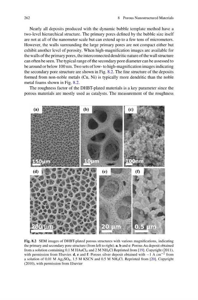
be around or below 100 nm. Two sets of low- to high-magnification images indicating
the secondary pore structure are shown in Fig. 8.2. The fine structure of the deposits
formed from non-noble metals (Cu, Ni) is typically more dendritic than the noble
metal foams shown in Fig. 8.2.
The roughness factor of the DHBT-plated materials is a key parameter since the
porous materials are mostly used as catalysts. The measurement of the roughness
Fig. 8.2 SEM images of DHBT-plated porous structures with various magnifications, indicating
the primary and secondary pore structure (from left to right). a, b and c: Porous Au deposit obtained
from a solution containing 0.1 M HAuCl 4 and 2 M NH 4 Cl Reprinted from [19]. Copyright (2011),
with permission from Elsevier. d, e and f: Porous silver deposit obtained with −1 A cm −2 from
a solution of 0.01 M Ag 2 SO 4 , 1.5 M KSCN and 0.5 M NH 4 Cl. Reprinted from [20]. Copyright
(2010), with permission from Elsevier
8 Porous Nanostructured Materials
Nearly all deposits produced with the dynamic bubble template method have a
two-level hierarchical structure. The primary pores defined by the bubble size itself
are not at all of the nanometer scale but can extend up to a few tens of micrometers.
However, the walls surrounding the large primary pores are not compact either but
exhibit another level of porosity. When high-magnification images are available for
the walls of the primary pores, the interconnected dendritic nature of the wall structure
can often be seen. The typical range of the secondary pore diameter can be assessed to
be around or below 100 nm. Two sets of low- to high-magnification images indicating
the secondary pore structure are shown in Fig. 8.2. The fine structure of the deposits
formed from non-noble metals (Cu, Ni) is typically more dendritic than the noble
metal foams shown in Fig. 8.2.
The roughness factor of the DHBT-plated materials is a key parameter since the
porous materials are mostly used as catalysts. The measurement of the roughness
Fig. 8.2 SEM images of DHBT-plated porous structures with various magnifications, indicating
the primary and secondary pore structure (from left to right). a, b and c: Porous Au deposit obtained
from a solution containing 0.1 M HAuCl 4 and 2 M NH 4 Cl Reprinted from [19]. Copyright (2011),
with permission from Elsevier. d, e and f: Porous silver deposit obtained with −1 A cm −2 from
a solution of 0.01 M Ag 2 SO 4 , 1.5 M KSCN and 0.5 M NH 4 Cl. Reprinted from [20]. Copyright
(2010), with permission from Elsevier