

38
therefore recommend that one initially perform a positive binding
test using saturating levels of ligand by means of a rapid and not
necessarily quantitative assay including size exclusion chromatography, affinity pull-down, or shift in T m or C m . If no binding is
observed, then one or more destabilizing mutations are introduced
into the N-fold, e.g., substituting Gly, Ala, or Val at one or more
hydrophobic packing sites identified in Subheading 3. A positive
binding result indicates that the protein has adopted the N′-fold in
the presence of ligand but does not reveal whether it had switched
to the N′-fold from the N-fold, or was already in the N′-fold in the
absence of ligand.
Once a binding interaction has been established, the final step is to
optimize the sensor for maximum fluorescence response and binding affinity. Fluorescent donor and acceptor groups are attached to
POI-AFF at its N-terminus and in the permutation loop of the
N-frame. We favor attaching maleimide dyes to engineered Cys
residues, although there are a variety of alternate chemistries and
labeling strategies from which to choose
2
. The important considerations are that one obtains close to a 1:1 donor:acceptor ratio and
that the chosen dyes are sensitive to short-range distance changes
(short Förster radii for FRET), as they will be close enough to be
in contact in the N′-fold and of variable distance apart in the
N-fold, depending on the length and residual structure of the
duplicated segment. To achieve 1:1 donor:acceptor labeling one
may have to experiment with different ratios of dyes in the labeling
step. We have also obtained satisfactory results by labeling both
positions with a single fluorophore (pyrene excimer formation and
BODIPY-FL self-quenching).
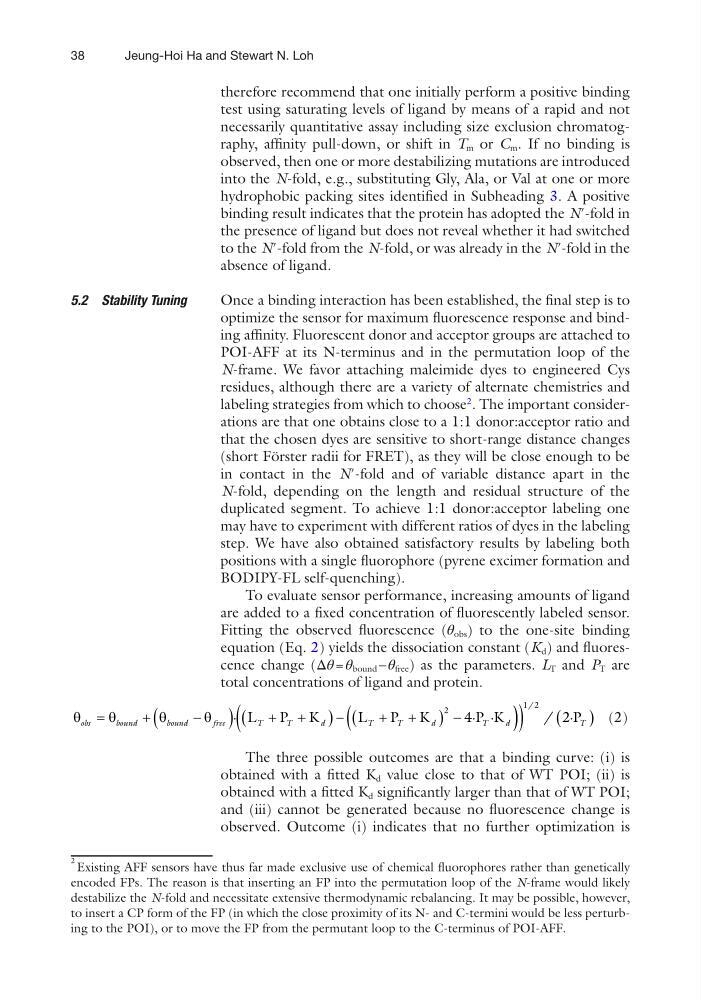
To evaluate sensor performance, increasing amounts of ligand
are added to a fixed concentration of fluorescently labeled sensor.
Fitting the observed fluorescence (θ obs ) to the one-site binding
equation (Eq. 2) yields the dissociation constant (K d ) and fluorescence change (Δθ = θ bound – θ free ) as the parameters. L T and P T are
total concentrations of ligand and protein.
q
q
q
q
obs
b ound
bound
free
T
T
d
T
T
d
T
d
=
+
-
(
) + +
(
)+ +
(
)(
·
· ·
L
P K
L
P K
P K
2
4
) )
(
) ( )
1 2
2
/ / ·P T
(2)
The three possible outcomes are that a binding curve: (i) is
obtained with a fitted K d value close to that of WT POI; (ii) is
obtained with a fitted K d significantly larger than that of WT POI;
and (iii) cannot be generated because no fluorescence change is
observed. Outcome (i) indicates that no further optimization is
2 Existing AFF sensors have thus far made exclusive use of chemical fluorophores rather than genetically
encoded FPs. The reason is that inserting an FP into the permutation loop of the N-frame would likely
destabilize the N-fold and necessitate extensive thermodynamic rebalancing. It may be possible, however,
to insert a CP form of the FP (in which the close proximity of its N- and C-termini would be less perturbing to the POI), or to move the FP from the permutant loop to the C-terminus of POI-AFF.
5.2 Stability Tuning
Jeung-Hoi Ha and Stewart N. Loh
therefore recommend that one initially perform a positive binding
test using saturating levels of ligand by means of a rapid and not
necessarily quantitative assay including size exclusion chromatography, affinity pull-down, or shift in T m or C m . If no binding is
observed, then one or more destabilizing mutations are introduced
into the N-fold, e.g., substituting Gly, Ala, or Val at one or more
hydrophobic packing sites identified in Subheading 3. A positive
binding result indicates that the protein has adopted the N′-fold in
the presence of ligand but does not reveal whether it had switched
to the N′-fold from the N-fold, or was already in the N′-fold in the
absence of ligand.
Once a binding interaction has been established, the final step is to
optimize the sensor for maximum fluorescence response and binding affinity. Fluorescent donor and acceptor groups are attached to
POI-AFF at its N-terminus and in the permutation loop of the
N-frame. We favor attaching maleimide dyes to engineered Cys
residues, although there are a variety of alternate chemistries and
labeling strategies from which to choose
2
. The important considerations are that one obtains close to a 1:1 donor:acceptor ratio and
that the chosen dyes are sensitive to short-range distance changes
(short Förster radii for FRET), as they will be close enough to be
in contact in the N′-fold and of variable distance apart in the
N-fold, depending on the length and residual structure of the
duplicated segment. To achieve 1:1 donor:acceptor labeling one
may have to experiment with different ratios of dyes in the labeling
step. We have also obtained satisfactory results by labeling both
positions with a single fluorophore (pyrene excimer formation and
BODIPY-FL self-quenching).
To evaluate sensor performance, increasing amounts of ligand
are added to a fixed concentration of fluorescently labeled sensor.
Fitting the observed fluorescence (θ obs ) to the one-site binding
equation (Eq. 2) yields the dissociation constant (K d ) and fluorescence change (Δθ = θ bound – θ free ) as the parameters. L T and P T are
total concentrations of ligand and protein.
q
q
q
q
obs
b ound
bound
free
T
T
d
T
T
d
T
d
=
+
-
(
) + +
(
)+ +
(
)(
·
· ·
L
P K
L
P K
P K
2
4
) )
(
) ( )
1 2
2
/ / ·P T
(2)
The three possible outcomes are that a binding curve: (i) is
obtained with a fitted K d value close to that of WT POI; (ii) is
obtained with a fitted K d significantly larger than that of WT POI;
and (iii) cannot be generated because no fluorescence change is
observed. Outcome (i) indicates that no further optimization is
2 Existing AFF sensors have thus far made exclusive use of chemical fluorophores rather than genetically
encoded FPs. The reason is that inserting an FP into the permutation loop of the N-frame would likely
destabilize the N-fold and necessitate extensive thermodynamic rebalancing. It may be possible, however,
to insert a CP form of the FP (in which the close proximity of its N- and C-termini would be less perturbing to the POI), or to move the FP from the permutant loop to the C-terminus of POI-AFF.
5.2 Stability Tuning
Jeung-Hoi Ha and Stewart N. Loh