

36
encodes for the N-terminal half of the linker, and ends with the
same restriction site as in Subheading 4.1. PCR amplify to generate
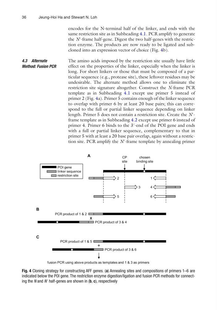
the N′-frame half-gene. Digest the two half-genes with the restriction enzyme. The products are now ready to be ligated and subcloned into an expression vector of choice (Fig. 4b).
The amino acids imposed by the restriction site usually have little
effect on the properties of the linker, especially when the linker is
long. For short linkers or those that must be composed of a particular sequence (e.g., protease site), these leftover residues may be
undesirable. The alternate method allows one to eliminate the
restriction site signature altogether. Construct the N-frame PCR
template as in Subheading 4.1 except use primer 5 instead of
primer 2 (Fig. 4a). Primer 5 contains enough of the linker sequence
to overlap with primer 6 by at least 20 base pairs; this can correspond to the full or partial linker sequence depending on linker
length. Primer 5 does not contain a restriction site. Create the N′frame template as in Subheading 4.2 except use primer 6 instead of
primer 4. Primer 6 binds to the 3′-end of the POI gene and ends
with a full or partial linker sequence, complementary to that in
primer 5 with at least a 20 base pair overlap, again without a restriction site. PCR amplify the N′-frame template by annealing primer
4.3 Alternate
Method: Fusion PCR
Fig. 4 Cloning strategy for constructing AFF genes. (a) Annealing sites and compositions of primers 1–6 are
indicated below the POI gene. The restriction enzyme digestion/ligation and fusion PCR methods for connecting the N and N′ half-genes are shown in (b, c), respectively
Jeung-Hoi Ha and Stewart N. Loh
encodes for the N-terminal half of the linker, and ends with the
same restriction site as in Subheading 4.1. PCR amplify to generate
the N′-frame half-gene. Digest the two half-genes with the restriction enzyme. The products are now ready to be ligated and subcloned into an expression vector of choice (Fig. 4b).
The amino acids imposed by the restriction site usually have little
effect on the properties of the linker, especially when the linker is
long. For short linkers or those that must be composed of a particular sequence (e.g., protease site), these leftover residues may be
undesirable. The alternate method allows one to eliminate the
restriction site signature altogether. Construct the N-frame PCR
template as in Subheading 4.1 except use primer 5 instead of
primer 2 (Fig. 4a). Primer 5 contains enough of the linker sequence
to overlap with primer 6 by at least 20 base pairs; this can correspond to the full or partial linker sequence depending on linker
length. Primer 5 does not contain a restriction site. Create the N′frame template as in Subheading 4.2 except use primer 6 instead of
primer 4. Primer 6 binds to the 3′-end of the POI gene and ends
with a full or partial linker sequence, complementary to that in
primer 5 with at least a 20 base pair overlap, again without a restriction site. PCR amplify the N′-frame template by annealing primer
4.3 Alternate
Method: Fusion PCR
Fig. 4 Cloning strategy for constructing AFF genes. (a) Annealing sites and compositions of primers 1–6 are
indicated below the POI gene. The restriction enzyme digestion/ligation and fusion PCR methods for connecting the N and N′ half-genes are shown in (b, c), respectively
Jeung-Hoi Ha and Stewart N. Loh