

259
recruitment toward the membrane leads to the activation of AKT1.
The validation of kinase activation is shown by Western blot analysis
using antibodies against AKT, AKT pT308 , and AKT pS473 . The used
constructs further contain fluorescence proteins, which further allow
the visualization of light- dependent membrane recruitment via fluorescence microscopy. The described methods can be transferred to
other protein kinases depending on oligomerization or recruitment
to defined subcellular regions for activation.
2 Materials
1. Culture medium: Dulbecco’s modified Eagle medium
(DMEM) supplemented with 10% (v/v) fetal calf serum (FCS)
and penicillin (100 U/ml)/streptomycin (100 μg/ml). Stored
at 4 °C.
2. Phosphate buffered saline (PBS): 10 mM Na 2 HPO 4 , 2 mM
KH 2 PO 4 , 137 mM NaCl, and 2.7 mM KCl, pH 7.4.
3. Trypsin-EDTA solution: 0.05% Trypsin, 0.02% EDTA in PBS
without Ca
2+
and Mg
2+
. Stored at 4 °C.
4. CASY model TT cell counting device, 150 μm (Roche, cat. no.
05651697001) with CASY cups (Roche, cat. no. 05651794001)
and CASY ton buffer (Roche, cat. no. 05651808001).
Alternatively, a standard hemocytometer can be used.
2.1 Cell Culture
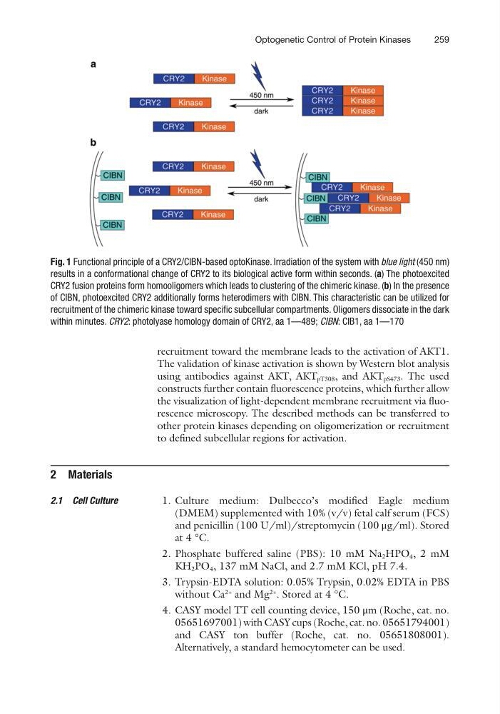
Fig. 1 Functional principle of a CRY2/CIBN-based optoKinase. Irradiation of the system with blue light (450 nm)
results in a conformational change of CRY2 to its biological active form within seconds. (a) The photoexcited
CRY2 fusion proteins form homooligomers which leads to clustering of the chimeric kinase. (b) In the presence
of CIBN, photoexcited CRY2 additionally forms heterodimers with CIBN. This characteristic can be utilized for
recruitment of the chimeric kinase toward specific subcellular compartments. Oligomers dissociate in the dark
within minutes. CRY2: photolyase homology domain of CRY2, aa 1—489; CIBN: CIB1, aa 1—170
Optogenetic Control of Protein Kinases
recruitment toward the membrane leads to the activation of AKT1.
The validation of kinase activation is shown by Western blot analysis
using antibodies against AKT, AKT pT308 , and AKT pS473 . The used
constructs further contain fluorescence proteins, which further allow
the visualization of light- dependent membrane recruitment via fluorescence microscopy. The described methods can be transferred to
other protein kinases depending on oligomerization or recruitment
to defined subcellular regions for activation.
2 Materials
1. Culture medium: Dulbecco’s modified Eagle medium
(DMEM) supplemented with 10% (v/v) fetal calf serum (FCS)
and penicillin (100 U/ml)/streptomycin (100 μg/ml). Stored
at 4 °C.
2. Phosphate buffered saline (PBS): 10 mM Na 2 HPO 4 , 2 mM
KH 2 PO 4 , 137 mM NaCl, and 2.7 mM KCl, pH 7.4.
3. Trypsin-EDTA solution: 0.05% Trypsin, 0.02% EDTA in PBS
without Ca
2+
and Mg
2+
. Stored at 4 °C.
4. CASY model TT cell counting device, 150 μm (Roche, cat. no.
05651697001) with CASY cups (Roche, cat. no. 05651794001)
and CASY ton buffer (Roche, cat. no. 05651808001).
Alternatively, a standard hemocytometer can be used.
2.1 Cell Culture
Fig. 1 Functional principle of a CRY2/CIBN-based optoKinase. Irradiation of the system with blue light (450 nm)
results in a conformational change of CRY2 to its biological active form within seconds. (a) The photoexcited
CRY2 fusion proteins form homooligomers which leads to clustering of the chimeric kinase. (b) In the presence
of CIBN, photoexcited CRY2 additionally forms heterodimers with CIBN. This characteristic can be utilized for
recruitment of the chimeric kinase toward specific subcellular compartments. Oligomers dissociate in the dark
within minutes. CRY2: photolyase homology domain of CRY2, aa 1—489; CIBN: CIB1, aa 1—170
Optogenetic Control of Protein Kinases