

150
and antigen-dependent growth-enhancement of E. coli expressing
the fusion protein in the presence of ampicillin. However, the
increase of the specific activity was only up to 25% and there was a
room for improvement.
More recently, aiming to transduce the antigen-dependent
binding signal more effectively to the catalytic domain, we tried to
optimize Fv-cpBLA by recombining the antibody domains to
cpBLA more directly instead of using commonly employed flexible
linkers [10]. The distance between the N- and C-termini of antibody variable domain V H and V L is between 30 and 40 Å. This is
considered too long and may disrupt the optimal orientation of
protein fragments or the conformation of circularly permuted
enzymes. Therefore, based on the previous report [11], we focused
on the 3 and 3b loops located far from antigen-binding loops but
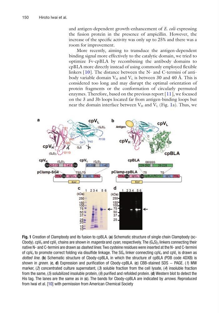
near the domain interface between V H and V L (Fig. 1a). Thus, we
Fig. 1 Creation of Clampbody and its fusion to cpBLA. (a) Schematic structure of single chain Clampbody (scCbody). cpV H and cpV L chains are shown in magenta and cyan, respectively. The (G 4 S) 3 linkers connecting their
native N- and C-termini are drawn as dashed lines. Two cysteine residues were inserted at the N- and C-termini
of cpV H to promote correct folding via disulfide linkage. The SG 4 linker connecting cpV H and cpV L is drawn as
dotted line. (b) Schematic structure of Cbody-cpBLA, in which the structure of cpBLA (PDB code 4DXB) is
shown in green. (c, d) Expression and purification of Cbody-cpBLA. (c) CBB-stained SDS − PAGE. (1) MW
marker, (2) concentrated culture supernatant, (3) soluble fraction from the cell lysate, (4) insoluble fraction
from the same, (5) solubilized insoluble protein, (6) purified and refolded protein. (d) Western blot to detect the
His tag. The lanes are the same as in (c). The bands for Cbody-cpBLA are indicated by arrows. Reproduced
from Iwai et al. [10] with permission from American Chemical Society
Hiroto Iwai et al.
and antigen-dependent growth-enhancement of E. coli expressing
the fusion protein in the presence of ampicillin. However, the
increase of the specific activity was only up to 25% and there was a
room for improvement.
More recently, aiming to transduce the antigen-dependent
binding signal more effectively to the catalytic domain, we tried to
optimize Fv-cpBLA by recombining the antibody domains to
cpBLA more directly instead of using commonly employed flexible
linkers [10]. The distance between the N- and C-termini of antibody variable domain V H and V L is between 30 and 40 Å. This is
considered too long and may disrupt the optimal orientation of
protein fragments or the conformation of circularly permuted
enzymes. Therefore, based on the previous report [11], we focused
on the 3 and 3b loops located far from antigen-binding loops but
near the domain interface between V H and V L (Fig. 1a). Thus, we
Fig. 1 Creation of Clampbody and its fusion to cpBLA. (a) Schematic structure of single chain Clampbody (scCbody). cpV H and cpV L chains are shown in magenta and cyan, respectively. The (G 4 S) 3 linkers connecting their
native N- and C-termini are drawn as dashed lines. Two cysteine residues were inserted at the N- and C-termini
of cpV H to promote correct folding via disulfide linkage. The SG 4 linker connecting cpV H and cpV L is drawn as
dotted line. (b) Schematic structure of Cbody-cpBLA, in which the structure of cpBLA (PDB code 4DXB) is
shown in green. (c, d) Expression and purification of Cbody-cpBLA. (c) CBB-stained SDS − PAGE. (1) MW
marker, (2) concentrated culture supernatant, (3) soluble fraction from the cell lysate, (4) insoluble fraction
from the same, (5) solubilized insoluble protein, (6) purified and refolded protein. (d) Western blot to detect the
His tag. The lanes are the same as in (c). The bands for Cbody-cpBLA are indicated by arrows. Reproduced
from Iwai et al. [10] with permission from American Chemical Society
Hiroto Iwai et al.