

132
optical imaging reporters to measure the kinase activity of various
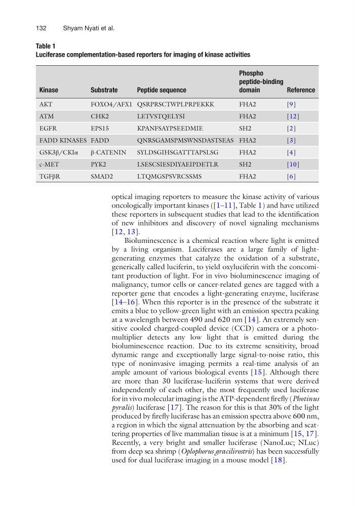
oncologically important kinases ([1–11], Table 1) and have utilized
these reporters in subsequent studies that lead to the identification
of new inhibitors and discovery of novel signaling mechanisms
[12, 13].
Bioluminescence is a chemical reaction where light is emitted
by a living organism. Luciferases are a large family of lightgenerating enzymes that catalyze the oxidation of a substrate,
generically called luciferin, to yield oxyluciferin with the concomitant production of light. For in vivo bioluminescence imaging of
malignancy, tumor cells or cancer-related genes are tagged with a
reporter gene that encodes a light-generating enzyme, luciferase
[14–16]. When this reporter is in the presence of the substrate it
emits a blue to yellow-green light with an emission spectra peaking
at a wavelength between 490 and 620 nm [14]. An extremely sensitive cooled charged-coupled device (CCD) camera or a photomultiplier detects any low light that is emitted during the
bioluminescence reaction. Due to its extreme sensitivity, broad
dynamic range and exceptionally large signal-to-noise ratio, this
type of noninvasive imaging permits a real-time analysis of an
ample amount of various biological events [15]. Although there
are more than 30 luciferase-luciferin systems that were derived
independently of each other, the most frequently used luciferase
for in vivo molecular imaging is the ATP-dependent firefly (Photinus
pyralis) luciferase [17]. The reason for this is that 30% of the light
produced by firefly luciferase has an emission spectra above 600 nm,
a region in which the signal attenuation by the absorbing and scattering properties of live mammalian tissue is at a minimum [15, 17].
Recently, a very bright and smaller luciferase (NanoLuc; NLuc)
from deep sea shrimp (Oplophorus gracilirostris) has been successfully
used for dual luciferase imaging in a mouse model [18].
Table 1
Luciferase complementation-based reporters for imaging of kinase activities
Kinase
Substrate
Peptide sequence
Phospho
peptide- binding
domain
Reference
AKT
FOXO4/AFX1 QSRPRSCTWPLPRPEKKK
FHA2
[9]
ATM
CHK2
LETVSTQELYSI
FHA2
[12]
EGFR
EPS15
KPANFSAYPSEEDMIE
SH2
[2]
FADD KINASES FADD
QNRSGAMSPMSWNSDASTSEAS FHA2
[3]
GSK3β/CKIα
β-CATENIN
SYLDSGIHSGATTTAPSLSG
FHA2
[4]
c-MET
PYK2
LSESCSIESDIYAEIPDETLR
SH2
[10]
TGFβR
SMAD2
LTQMGSPSVRCSSMS
FHA2
[6]
Shyam Nyati et al.
optical imaging reporters to measure the kinase activity of various
oncologically important kinases ([1–11], Table 1) and have utilized
these reporters in subsequent studies that lead to the identification
of new inhibitors and discovery of novel signaling mechanisms
[12, 13].
Bioluminescence is a chemical reaction where light is emitted
by a living organism. Luciferases are a large family of lightgenerating enzymes that catalyze the oxidation of a substrate,
generically called luciferin, to yield oxyluciferin with the concomitant production of light. For in vivo bioluminescence imaging of
malignancy, tumor cells or cancer-related genes are tagged with a
reporter gene that encodes a light-generating enzyme, luciferase
[14–16]. When this reporter is in the presence of the substrate it
emits a blue to yellow-green light with an emission spectra peaking
at a wavelength between 490 and 620 nm [14]. An extremely sensitive cooled charged-coupled device (CCD) camera or a photomultiplier detects any low light that is emitted during the
bioluminescence reaction. Due to its extreme sensitivity, broad
dynamic range and exceptionally large signal-to-noise ratio, this
type of noninvasive imaging permits a real-time analysis of an
ample amount of various biological events [15]. Although there
are more than 30 luciferase-luciferin systems that were derived
independently of each other, the most frequently used luciferase
for in vivo molecular imaging is the ATP-dependent firefly (Photinus
pyralis) luciferase [17]. The reason for this is that 30% of the light
produced by firefly luciferase has an emission spectra above 600 nm,
a region in which the signal attenuation by the absorbing and scattering properties of live mammalian tissue is at a minimum [15, 17].
Recently, a very bright and smaller luciferase (NanoLuc; NLuc)
from deep sea shrimp (Oplophorus gracilirostris) has been successfully
used for dual luciferase imaging in a mouse model [18].
Table 1
Luciferase complementation-based reporters for imaging of kinase activities
Kinase
Substrate
Peptide sequence
Phospho
peptide- binding
domain
Reference
AKT
FOXO4/AFX1 QSRPRSCTWPLPRPEKKK
FHA2
[9]
ATM
CHK2
LETVSTQELYSI
FHA2
[12]
EGFR
EPS15
KPANFSAYPSEEDMIE
SH2
[2]
FADD KINASES FADD
QNRSGAMSPMSWNSDASTSEAS FHA2
[3]
GSK3β/CKIα
β-CATENIN
SYLDSGIHSGATTTAPSLSG
FHA2
[4]
c-MET
PYK2
LSESCSIESDIYAEIPDETLR
SH2
[10]
TGFβR
SMAD2
LTQMGSPSVRCSSMS
FHA2
[6]
Shyam Nyati et al.